HoldingOrders.com

Altered Mental Status

Source: Olsen, Alexander. (2014). Cognitive Control Function and Moderate-to-Severe Traumatic Brain Injury: Functional and Structural Brain Correlates.

Source: Olsen, Alexander. (2014). Cognitive Control Function and Moderate-to-Severe Traumatic Brain Injury: Functional and Structural Brain Correlates.

Trauma

  • Concussion: If no loss of consciousness, decreased responsiveness, repetitive vomiting, seizure, and/or focal neurologic deficits →

    • Do not obtain MRI

    • Initiate 6-day graded return to play: Regular activities (school) → light aerobic activity → moderate activity → heavy, non-contact → practice with full contact → competition

  • Epidural hematoma

    • Adolescent s/p head trauma and a lucid interval presenting with acute onset altered consciousness, headache, vomiting, aphasia, seizures, unilateral weakness

    • Head CT shows lens-shaped collection of epidural blood

    • Consult surgery and do not administer glucocorticoids

    • Mortality 5 to 10 percent

  • Glasgow Coma Scale < 8: Intubate

Focal Neurologic Deficit and/or Convulsions

Syncope (Transient Cerebral Hypoperfusion)

Syncope.jpg

Differential Diagnosis

  • Neurally mediated (45%): Increased parasympathetic/vagal tone → bradycardia and hypotension

    • Vasovagal: Warm/crowded space, prolonged standing, emotion/fear/pain, nausea, transient diaphoresis

    • Situational: S/p coughing, voiding after a meal

    • Consider seizure (eyes open) and psychogenic syncope (eyes closed)

  • Cardiac (20%)

    • Arrhythmia (most common): Elderly patient with h/o atrial fibrillation/flutter and family h/o sudden/unexplained death presents with palpitations, abnormal EKG. Treatment per arrhythmia identified.

    • Structural cardiac abnormality/cardiomyopathy: Elderly, h/o valvular or infiltrative (sarcoidosis, hemochromatosis, amyloidosis) heart disease presents with chest pain at rest, syncope during exercise. Evidence of heart failure on physical exam and PR interval > 200 ms (heart block) on EKG.

    • Hypertrophic cardiomyopathy: Pediatric patient with family h/o sudden cardiac death presents with new onset syncope while exercising in hot weather. Start nadolol and transition to verapamil if initial treatment is ineffective.

  • Orthostatic hypotension (10%): Decrease of ≥ 20 mmHg systolic or ≥ 10 mmHg diastolic within 3 minutes of moving from supine to upright position

    • Patient with h/o autonomic dysfunction due to alcoholism, DM, Parkinson disease, multiple sclerosis presents with syncope during postural change. Recent, acute volume loss due to dehydration, hemorrhage. Consumes low-salt diet.

    • Recent changes in medications: Alpha blockers (tamsulosin), beta-blockers (metoprolol), calcium channel blockers (amlodipine), diuretics (furosemide), phosphodiesterase inhibitors (e.g. sildenafil), SSRIs

    • Tachycardia and positive orthostatic vital signs on exam

Evaluation

  • Obtain history with focus on precipitating events, h/o cardiac disease, and clinical features (italicized) above

  • See section on syncope

Delirium, Encephalopathy, Confusion

Dementia

  • Alzheimer's disease (review medications, behavioral intervention, aripiprazole)

  • Lewy body dementia

  • Vascular dementia

  • Frontotemporal dementia

Anxiety/Depression

Anxiety

Episodic

Social Anxiety Disorder

  • Anxiety correlated with specific social and/or cultural interactions

  • Refer for cognitive behavioral therapy and/or graded desensitization

  • Start escitalopram 10 mg qd and propranolol 40 mg PRN 60 minutes prior to anxiety-provoking events

  • Buspirone is not effective

Panic Disorder

  • Acute onset anxiety presenting with lightheadedness, shortness of breath

  • Hyperventilation, diaphoresis, and tremors on exam

  • Treatment

    • Start escitalopram 10 mg qd and hydroxyzine 25 mg TID PRN

      • Continue escitalopram for at least 1 year before trial of discontinuation

      • Increase escitalopram by 10 mg every 4 weeks until symptoms are controlled (maximum daily dose 30 mg qd)

    • Buspirone is not effective

Chronic

Generalized Anxiety Disorder

  • More common in females (2:1 prevalence)

  • Patients often perseverate on personal failures and imperfections

  • Often comorbid with major depressive disorder

  • Diagnosis

    • Excessive anxiety and worry (apprehensive expectation), occurring more days than not for at least 6 months

    • The individual finds it difficult to control the worry

    • The anxiety and worry are associated with three (or more) of the following six symptoms

      • Restlessness, feeling keyed up or on edge

      • Being easily fatigued

      • Difficulty concentrating or mind going blank

      • Irritability

      • Muscle tension

      • Sleep disturbance (difficulty falling or staying asleep, or restless, unsatisfying sleep)

    • The anxiety, worry, or physical symptoms cause clinically significant distress or impairment in social, occupational, or other important areas of functioning

    • The disturbance is not attributable to the physiological effects of a substance (e.g. substance abuse) or another medical condition (e.g. hyperthyroidism)

    • The disturbance is not better explained by another medical disorder (e.g. panic disorder)

  • Treatment

    • Maintenance medications: Start one of the following

      • Escitalopram 10 mg qd and increase by 10 mg every 4 weeks until symptoms are controlled (maximum daily dose 30 mg qd)

      • Buspirone 5 mg BID and increase 5 mg BID every week until symptoms are controlled (maximum daily dose 20 mg TID)

    • Consider hydroxyzine 25 mg TID PRN for comorbid panic attacks

    • Beta-blockers (e.g. propranolol) are not effective

Due to a medical condition: Hyperthyroidism

Major and Persistent Depression Disorders

Overview

  • Screen all patients ≥ 12 years old

  • Organic etiologies

    • Associated medical conditions commonly associated with fatigue

    • Review social history and medications for central nervous system depressants, e.g.

      • Alcohol and/or recreational drug use

      • Prescription anxiolytics, sedatives, narcotics

      • Corticosteroids

    • Consider CBC, CMP, TSH, RPR, urine drug screen prior to starting treatment

Diagnosis per DSM-5

Major Depressive Disorder

  • Diagnosis (PHQ-9) requires ≥ 5 of the following symptoms and symptoms 1 or 2 (below) must be present:

  1. Anhedonia (diminished interest or pleasure)

  2. Depressed mood most of the day, nearly every day

  3. Insomnia or hypersomnia

  4. Fatigue and/or decreased energy

  5. Poor appetite or overeating

  6. Feelings of worthlessness or excessive guilt

  7. Diminished concentration and/or decisiveness

  8. Slowing in thought or movement noticeable to others

  9. Recurrent thoughts of death or suicidal ideation without a plan

Persistent Depressive Disorder (Dysthymia)

  • Depressed mood for ≥ 2 years with no symptom free period for > 2 months and 2 of the following

    • Changes in sleep or appetite

    • Decreased energy or concentration

    • Slowing in thought or movement noticeable to others

    • Excessive guilt or thoughts of death

    • Hopelessness

Pseudodementia

  • Major depressive disorder in elderly patients leading to significant memory/concentration deficits patients

  • Resolves with treatment of depression

Treatment: Psychotherapy and/or Pharmacotherapy

  • Medications

    • First line: SSRI

      • Start escitalopram 10 mg qd and increase by 10 mg every 4 weeks until symptoms are controlled (maximum daily dose 30 mg qd)

      • Use caution in patients with a h/o mania, hyponatremia

    • Second line: SNRIs

      • Venlafaxine 75 mg qd

      • History of chronic pain: Duloxetine 40 mg qd

    • Sexual side effects with SSRI/SNRI and/or current smoker: Bupropion 150 mg qd (verify no h/o seizure)

    • Insomnia and

      • Decreased appetite: Mirtazapine 15 mg qhs

      • Chronic pain: Nortriptyline 50 mg qd

    • Refractory to multiple medications: Consider electroconvulsive therapy (ECT)

  • Schedule follow-up in 4-6 weeks

    • If repeat PHQ-9 shows improvement, continue current medication and consider increasing the dose

    • If no improvement, transition to another antidepressant

  • Duration of antidepressant treatment

    • First depressive episode: At least 6 months

    • Recurrent episode: At least 2 years

Depression-Associated Disorders

General Population

Seasonal Affective Disorder

  • Sub-category of other depressive disorders, e.g. MDD, bipolar

  • Diagnosis

    • Seasonal onset and remission of depressive, manic, or hypomanic episodes with

      • At least two seasonal depressive episodes within the last two years

      • More seasonal than non-seasonal depressive episodes

    • Not due to

      • Psychosocial stressors associated with a season, e.g. financial stress during the holidays

      • Another medical issue, e.g. hypothyroidism, substance abuse

  • Effective treatments: Light therapy (remain 12-18 inches from 10,000 lux lamp for 30 minutes/day), SSRI, CBT

Bipolar I and II

  • Diagnosis

    • Bipolar I: One manic episode lasting ≥ 1 week

    • Bipolar II: One hypomanic episode lasting ≥ 4 days and one major depressive episode (no history of manic episodes)

    • Manic episode (DIG FAST): Distractibility, indiscretion, grandiosity, flight of ideas, activity increase, sleep deficit, talkativeness (pressured speech)

    • Hypomanic episode: Less severe than a manic episode (socially appropriate, no long-lasting self harm, no need for hospitalization)

  • Treatment

    • Lithium (salt that adversely affects pregnancy, renal function)

      • Obtain initial baseline BMP, TSH, T4, and EKG and repeat q6 months

      • 300 mg TID and obtain lithium level within 5 days of starting mediation; goal 1.0 +/- 0.2 mEq/L

    • Valproic acid (anti-convulsant)

      • Obtain CMP (hepatotoxicity) and start 30 mg/kg/day

      • Adjust every 3 days (maximum daily dose 60 mg/kg/day) with goal serum level 50-100 mcg/mL

    • Quetiapine (Seroquel)

      • Start 100 mg BID and increase by 100 mg daily (maximum daily dose 800 mg)

      • Obtain yearly lipid panel, HbA1c

OBGYN-Related Conditions

  • Premenstrual syndrome and dysphoric disorder

  • Major depression disorder during pregnancy

    • Diagnosed per MDD criteria (see above)

    • Start or continue SSRI after shared decision making concerning medication safety

      • Safe: Escitalopram, sertraline

      • Possibly safe: Fluoxetine, venlafaxine, duloxetine, bupropion, amitriptyline

      • Contraindicated: Paroxetine, nortriptyline

  • Postpartum major depression (PMD)

    • Onset within 3 months of delivery with most cases (67%) starting within 6 weeks postpartum

    • Affects 10-15% of women

    • Risk factors

      • H/o major depressive disorder

      • Perinatal stress/trauma

      • Inadequate social support

    • Screen using Edinburgh Postnatal Depression Scale, if positive (score > 10):

      • Obtain TSH to rule out hypothyroidism

      • Start psychotherapy and/or fluoxetine 20 mg qd

Pediatric

  • Depression

    • Screen children 12 years and older using PHQ-9

    • Fluoxetine is the only FDA approved medication

  • Bipolar disorder: Common in children of patients with bipolar disorder

Depression and Anxiety Assessment

PHQ-2 for Depression

  • Over the last 2 weeks, how often have you been bothered by the following problems?

    • Little interest or pleasure in doing things?

    • Feeling down, depressed, or hopeless?

  • Responses

    • 0: Not at all

    • 1: Several days

    • 2: More than half of the days

    • 3: Nearly every day

  • Administer PHQ-9 for total score ≥ 3

Additional Questions Included in PHQ-9 for Depression

  • Trouble falling or staying asleep, or sleeping too much?

  • Feeling tired or having little energy?

  • Poor appetite or overeating?

  • Feeling bad about yourself — or that you are a failure or have let yourself or your family down?

  • Trouble concentrating on things, such as reading the newspaper or watching television?

  • Moving or speaking so slowly that other people could have noticed? Or so fidgety or restless that you have been moving a lot more than usual?

  • Thoughts that you would be better off dead, or thoughts of hurting yourself in some way?

GAD-7 for Anxiety

  • Over the last 2 weeks, how often have you been bothered by the following problems?

    • Feeling nervous, anxious or on edge

    • Not being able to stop or control worrying

    • Worrying too much about different things

    • Trouble relaxing

    • Being so restless that it is hard to sit still

    • Becoming easily annoyed or irritable

    • Feeling afraid as if something awful might happen

  • Responses

    • 0: Not at all

    • 1: Several days

    • 2: More than half of the days

    • 3: Nearly every day

  • Scoring

    • ≥ 5: Mild

    • ≥ 10: Moderate (probable GAD)

    • ≥ 15: Severe

Bleeding

GI Bleeding

Upper GI Bleeding: Melena → EGD before Colonoscopy

  • General management

    • Limit bleeding risk

      • Stop aspirin and NSAIDs

      • Reverse anticoagulants if possible, but do not delay EGD due to anticoagulation

    • Transition to NPO and start PPI IV (pantoprazole 80 mg loading dose followed by 40 mg q12 hours)

    • Transfuse for hemoglobin ≤ 7 mg/dL

    • Perform EGD within 24 hours of diagnosis

    • Glasgow-Blatchford score > 0: Hospitalize for 72 hours following EGD due to risk for rebleeding

  • Differential diagnosis

Lower GI Bleeding: Bright Red Blood per Rectum

  • Red flags requiring colonoscopy

    • Age ≥ 50 years

    • Family history of colon cancer, familial polyposis

    • Symptoms

      • Fever, unintentional weight loss

      • Change in stool frequency, caliber, consistency

    • Anemia, positive FOBT

  • Differential diagnosis

Etiology Prevalence by Age. Source: Pinar Solmaz Hasdemir, Mustafa Ulusoy, Esat Calik, Ulas Solmaz and Tevfik Guvenal (2016). Assessment of Patients with Abnormal Uterine Bleeding in the Reproductive Period According to the PALM-COEIN Classification…

Etiology Prevalence by Age. Source: Pinar Solmaz Hasdemir, Mustafa Ulusoy, Esat Calik, Ulas Solmaz and Tevfik Guvenal (2016). Assessment of Patients with Abnormal Uterine Bleeding in the Reproductive Period According to the PALM-COEIN Classification System

Abnormal Uterine Bleeding

Differential (PALM-COEIN)

  • Structural

  • Non-structural

Bleeding in Pregnancy

General Considerations

  • Light bleeding at 3-5 WGA is most likely due to implantation

  • Rule out cervical bleeding due to gonorrhea/Chlamydia

Early Pregnancy Bleeding (Algorithm)

  • Gestational trophoblastic disease

  • Ectopic pregnancy

  • Subchorionic hemorrhage

Late Pregnancy Bleeding

  • Placenta previa

  • Placental abruption

  • Vasa previa

Postpartum hemorrhage

Other

Von Willebrand Disease

  • Mucosal bleeding, e.g. menorrhagia

  • Most suggestive finding = prolonged PTT

  • Refer to hematologist for diagnosis confirmation 

Breast Issues

Breast Pain (Mastalgia)

  • Benign lactation complications

    • Severe engorgement: Encourage regular breast emptying

    • Obstructed milk duct

      • Presents as painful, non-erythematous, palpable lump

      • Recommendations include complete breast emptying q3h while awake

    • Galactocele

      • Milk retention cyst diagnosed with ultrasound, mammogram, or aspiration

      • Aspiration revealing milk is diagnostic and therapeutic

      • Consider referral to breast surgeon for recurrent cases

  • Mastitis

  • Breast abscess

  • Inflammatory breast cancer

Nipple Discharge

  • Pathologic, i.e. spontaneous, unilateral, and/or bloody: Refer to a breast surgeon

  • Non-bloody: Galactorrhea

Breast Mass

  • Fibroadenoma

  • Fibrocystic breast

  • Intraductal papilloma

  • Lobular carcinoma in situ

Cough

Adults

Infectious

Non-infectious

  • Acute cough

  • Chronic cough (duration ≥ 8 weeks)

    • Upper airway cough syndrome (post-nasal drip): Rhinorrhea, congestion, swollen turbinates, posterior pharyngeal cobblestoning

      • Allergic rhinitis: Start mometasone 50 mcg/spray BID +/- loratadine 10 mg daily and follow-up in 4 weeks

      • Non-allergic rhinitis: Start intranasal ipratropium bromide (0.03% solution) two sprays in each nostril BID and follow-up in 4 weeks

      • If no improvement at follow-up: Consider alternate etiology and treat accordingly. If concern for chronic rhinosinusitis, schedule sinus CT. Otherwise, refer to ENT for flexible sigmoidoscopy.

    • Asthma-induced cough

    • Non-asthmatic eosinophilic bronchitis

    • GERD-induced cough

    • ACE-inhibitor induced

    • Tobacco use

    • Less common etiologies: Pertussis, OSA, COPD, bronchiectasis, sarcoidosis

Hemoptysis

Etiologies

Initial workup: Obtain CXR

  • CXR shows infiltrate: Diagnose pneumonia and start azithromycin 500 mg on day 1 followed by 250 mg on days 2 through 5

  • Negative CXR

    • Low risk patient: No further treatment

    • High risk patient, i.e. age ≥ 40 years or ≥ 30 pack year smoking history: Obtain CT

      • Positive CT: Treatment per pathology

      • Negative CT: Consult pulmonology and consider bronchoscopy

Pediatrics

Constipation

Adult

Approach

  1. Red flags for colon cancer?

    • Sudden onset, severe constipation: FOBT and refer for colonoscopy if positive

    • Painless colorectal bleeding

      • Explore alternative diagnoses, e.g. anal fissure, hemorrhoids, anoreceptive intercourse

      • Refer for colonoscopy if age > 30 years and any of the following are true:

        • No alternative explanation

        • Persistent bleeding despite adequate treatment of alternative diagnosis

  2. Review risk factors and consider

    • Discontinuing offending medications

    • Obtaining HbA1c, TSH, UDS

  3. Treatment

    1. Trial of laxatives

    2. Laxatives ineffective

      • Initial test: Anorectal manometry and balloon expulsion test

      • Initial testing normal or inconclusive: Barium or MR defecography

Differential Diagnosis

Pediatric

Diarrhea

Bristol Stool Chart. Source: Cabot Health

Bristol Stool Chart. Source: Cabot Health

Consistency

Watery

  • Infectious: Acute gastroenteritis

    • Acutely increased stool frequency/volume, vomiting, infectious exposure, dry mucous membranes, hyperactive bowel sounds

    • Fecal lactoferrin, stool culture, stool ova and parasites, C. difficile toxin

    • Oral rehydration, BRAT diet, loperamide (non-bloody), Pepto-Bismol (bloody), simethicone, lactobacillus, antibiotics

  • Irritable bowel syndrome

    • Cramping abdominal pain with change in stool frequency/consistency for 2 to 3 months

    • CBC, TSH, CRP, ESR, FOBT, anti-tissue transglutaminase IgA, EGD

  • Microscopic colitis: Elderly patients. Increased risk with > 6 months daily NSAID use. Confirm with colon biopsy.

  • Laxative abuse

Fatty (malabsorption): Steatorrhea with gas +/- weight loss

Inflammatory: Irritable Bowel Disease

Additional Considerations

Dysphagia

Overview

Definitions

  • Dysphagia: Difficulty swallowing

  • Odynophagia: Painful swallowing that may be associated with dysphagia.

    • May be associated with dysphagia.

    • Consider EGD to evaluate for

      • Esophagitis (e.g. eosinophilic)

      • Post-surgical complications

Evaluation: Barium swallow → EGD → manometry

  • Concern for aspiration (e.g. s/p stroke): Obtain video fluoroscopy before barium swallow

  • Barium swallow assess motility and reveals obstructive lesions

  • EGD detects inflammation/erosion/infection and allows for biopsy

  • Manometry may reveal motility disorder missed on barium swallow

Motility Disorders (Solids + Liquids)

  • Sudden onset: Stroke-related deficits

  • Progressive symptoms: Neuromuscular motility disorder

    • Esophageal motility disorders (most common)

      • Esophageal spasm: Omeprazole 20 mg qd for GERD and peppermint oil for dysmotility. Diltiazem 60 mg q6h for refractory symptoms. Consider GI referral.

      • Scleroderma: Chronic heartburn, Renaud’s phenomenon, bloating, and alternating diarrhea/constipation. Skin thickening on fingers, hands, and face. Interstitial lung disease on CT chest. Obtain CBC, CMP, CK, U/A, ANA, anti-Scl-70 antibody, anticentromere antibody, and anti-RNA polymerase III antibody if suspected. Start omeprazole 20 mg BID for GERD. Trial of metoclopramide for refractory dysphagia symptoms. Refer to rehumatology for immunosuppressive therapy.

      • Achalasia: Frequent regurgitation and symptoms that improve with repeat swallowing

    • Oropharyngeal dysphagia due to neuromuscular disease

Mechanical Obstruction (Solids Only)

  • Pediatric or history of mental illness: Rule out foreign body ingestion

  • Intermittent symptoms

    • Esophageal web

    • Esophageal ring (Schatzki ring)

    • Zenker’s diverticulum: Halitosis with regurgitations of old food

  • Progressive symptoms

    • Stricture

      • Peptic

      • Radiation

      • Medications: NSAIDs, bisphosphonates, antibiotics (tetracyclines and sulfa derivatives)

    • Extra-esophageal compression

      • Left atrial enlargement

      • Vascular compression

      • Mediastinal mass

    • Malignancy: Patient age > 50 years. Weight loss and no regurgitation (as compared to achalasia).

Dyspnea

Pulmonary

Common Etiologies

Additional Considerations

  • Emergent/urgent

  • Pleural effusion

  • Interstitial lung disease

    • Infectious

      • Risk factors: Age > 60 years with h/o COPD, renal failure, and/or sepsis at presentation

      • Workup progression: Laboratory testing + CXR → chest CT → bronchoscopy with lavage → surgical biopsy (VATS)

    • Non-infectious

      • Pneumonia: Eosinophilic, hypersensitivity

      • Inflammatory/fibrotic: Interstitial pulmonary fibrosis, connective tissue disorders (e.g. scleroderma), systemic vasculitis

      • Exposure: Occupational, medications (e.g. nitrofurantoin or amiodarone), chemical, radiation (e.g. radiation pneumonitis)

CT Findings for Pulmonary Disease

  • Organizing pneumonia: Consolidation, nodules, perilobular pattern

  • Non-specific interstitial pneumonia: Traction bronchiectasis, reticulation, ground glass

  • Hypersensitivity pneumonitis: Traction bronchiectasis, honeycombing, centrilobular nodules, air trapping

  • Idiopathic pulmonary fibrosis: Traction bronchiectasis, honeycombing, reticulation

  • Sarcoid: Nodes, perilymphatic nodules

Cardiac

Other

  • Anxiety/Panic disorder

  • Metabolic acidosis

  • Neuromuscular

Dysuria

Edema

Acute Onset

Chronic

  • Cardiovascular (hydrostatic)

    • Heart failure

    • Aortic stenosis: Obtain echocardiogram and evaluate for

      • Congenital abnormality (e.g. bicuspid valve)

      • Rheumatic valve disease

      • Chronic valve calcification

    • Venous insufficiency

    • Pelvic compression (e.g. neoplasm): Abdominal U/S vs. CT

  • Hepatorenal (oncotic +/- hydrostatic)

Eye and Vision Disorders

Periocular Lesions

  • Urgent/Emergent

    • Preseptal/orbital cellulitis

    • Herpes zoster

  • Common

    • Affecting sebaceous glands: Blepharitis, stye, chalazion

    • Papular/nodular: Milia, xanthelasma, skin tag (acrochordons), seborrheic keratosis

    • Malignant: Basal cell carcinoma, squamous cell carcinoma

  • Additional considerations

    • Allergic reaction: Atopic dermatitis, urticaria +/- angioedema

    • Autoimmune: Heliotrope rash, vitiligo

    • Infectious: Verrucous lesions, molluscum

By https://www.scientificanimations.com - https://www.scientificanimations.com/wiki-images/, CC BY-SA 4.0, https://commons.wikimedia.org/w/index.php?curid=87227293

By https://www.scientificanimations.com - https://www.scientificanimations.com/wiki-images/, CC BY-SA 4.0, https://commons.wikimedia.org/w/index.php?curid=87227293

Red Eye

Differential Diagnosis

  • Trauma: Emergent ophthalmology referral in cases of moderate/severe pain, decreased visual acuity

    • Corneal abrasion

    • Foreign body

    • Chemical exposure

  • Vision changes: Emergent ophthalmology referral, especially if associated with pain

    • Keratitis

      • Corneal inflammation due to infection (bacteria, HSV, VZV, EBV, CMV), contact lenses, or corneal abrasion

      • Symptoms: Painful diminished vision +/- photophobia, foreign body sensation, mucopurulent discharge

      • Fluorescein/Wood’s lamp: Eyelid edema, hypopyon (leukocytes in anterior chamber), corneal ulcer

    • Iritis

      • Iris infection/inflammation due to perforating wound, corneal ulcer, or certain autoimmune conditions

      • Acute onset (hours) eye pain radiating to forehead/temple, diminished/blurred vision, photophobia

      • Eye watering/redness, ciliary/perilimbal injection, constricted/sluggish pupils

    • Acute angle-closure glaucoma

      • Obstructed aqueous humor outflow resulting in increased intraocular pressure

      • Acute onset severe/throbbing pain, diminished vision, light halos

      • Diffuse eye redness/watering, decreased visual acuity, pupil dilation with decreased reactivity to light, eye pain/firmness with palpation

  • Non-emergent

    • Blepharitis: Inflammatory condition of eyelid margins

    • Conjunctivitis (most common)

      • Allergic: Azelastine 0.05% (H1 receptor antagonist) 1 drop q12h

      • Keratoconjunctivitis (dry eye): Review history (Sjogren risk factors), medications (anticholinergic, antihistamine, OCPs) and consider trial of artificial tears

      • Viral: Symptomatic treatment

      • Bacterial: Erythromycin 0.5%, apply 0.5 inch long ribbon q6h x 7 days

    • Subconjunctival hemorrhage: Benign and resolve spontaneously

    • Episcleritis: Episcleral inflammation often associated with autoimmune disease (e.g. rheumatoid arthritis)

      • Generally self-limited with resolution in 3 weeks

      • Consider topical steroids for severe cases

Red Eye Ophthalmology Referrals

  • Emergent

    • Mechanism: Acute eye trauma

    • Symptoms

      • Moderate to severe pain

      • Acute vision loss: Associated with recent ocular surgery or concern for keratitis, iritis, acute angle closure glaucoma

    • Physical Exam

      • Vesicular rash (herpetic keratitis)

      • Severe mucopurulent discharge (hyperacute bacterial conjunctivitis)

      • Evidence corneal involvement with fluorescein/Wood's lamp

      • Distorted pupil

  • Non-emergent

    • History of recurrent eye infections or episcleritis episodes

    • Exam: Copious purulent discharge not improving with antibiotics

    • Treatment

      • Pain not relieved with topical anesthetic

      • Indication for starting topical steroids (e.g. severe episcleritis)

Vision Abnormalities/Loss

Adult

Acute: Require emergent evaluation by ophthalmology

  • Trauma: Physical and chemical injuries

  • Acute angle closure glaucoma

  • Retinal tear/detachment

  • Retinal vascular occlusion

    • Risk factors: HTN, DM

    • Sudden, painless visual loss

    • Tortuous, dilated retinal veins on fundoscopic exam +/- cotton wool spots, afferent pupillary defect on affected side

  • Optic nerve disease

Additional resource: Vision Loss in Older Persons, Fig. 2

Chronic

  • Emergent: Temporal (giant cell) arteritis

    • Epidemiology: Most common ages 70 - 79 years in patient with history of polymyalgia rheumatica

    • Presentation: Age > 50 years with new onset unilateral headache/visual disturbances +/- jaw claudication, fever, anemia, elevated ESR and/or CRP

    • Start prednisone 1 mg/kg (maximum 60 mg/day) q24h

    • Refer for color doppler ultrasonography vs. temporal artery biopsy (gold standard)

    • Consider additional screening for thoracic and abdominal aortic aneurysms due to increased risk

  • Age-related cataracts (most common)

    • Age > 40 years

    • Vision loss associated with glare intolerance/halos worse at night or while driving

    • Refer for surgical intervention when affecting activities of daily living

  • Diabetic retinopathy

    • Age > 50 years and more common in Hispanics

    • Fluctuating vision with floaters, light flashes, visual field deficits

    • Tight glycemic control, fenofibrate, panretinal photocoagulation

  • Glaucoma

    • Age > 40 years and more common in African Americans

    • Progressive peripheral and central field visual loss

    • Lower intraocular pressure with medicated eye drops vs. laser trabeculoplasty

  • Macular degeneration

    • Vision loss worse at night with positive Amsler grid test

    • Macular drusen and retinal pigment abnormalities

    • Slow progression with

      • Smoking cessation

      • AREDS2 vitamin supplementation to slow progression: Vitamin C 500 mg, vitamin E 400 IU, 25 mg zinc, 2 mg cupric oxide, 10 mg lutein, 2 mg zeaxanthin (e.g. PreserVision)

    • VEGF injections e.g. bevacizumab (Avastin) for neovascular subtype

Pediatric

Acute: Emergent consultation for trauma, orbital cellulitis, central retinal artery occlusion, and endophthalmitis

  • Painful

    • Trauma: Globe rupture, corneal injury, hyphema, lens dislocation, vitreous hemorrhage, retinal detachment, optic nerve injury

    • Surrounding skin changes: Orbital cellulitis

    • Red eye

      • Normal IOP: Keratoconjunctivitis (HSV vs. adenovirus)

      • Endophthalmitis (internal eye infection): Emergent consultation for possible intravitreal treatment and/or vitrectomy

      • Uveitis

    • Otherwise normal appearance

      • Painful EOM: Optic neuritis

      • Painless EOM: Ocular migraine

  • Painless

    • Leukorrhea present: Cataract vs. intraocular tumor

    • Retinal detachment/hemorrhage: Evaluate for child abuse

    • Central retinal artery occlusion: Consider in children with sickle cell disease

    • Optic migraine (non-emergent)

Chronic

  • Strabismus: Risk factor for visual loss

    • Unequal alignment and may be esotropic (adduction) or exotropic (abduction)

    • High risk for visual loss (see amblyopia)

    • Refer to pediatric ophthalmology with consideration for surgery if conservative treatment fails

  • Amblyopia

    • Reduction in visual acuity and may be strabismic (unequal alignment), refractive (unequal focus), or deprivational (structural abnormalities obscuring incoming image)

    • Treatment options: Patching, optical penalization, atropine eye drops

Facial Droop

Etiology Uncertain (i.e. Bell’s Palsy):

  • Start valacyclovir 1000 mg TID x 1 week + prednisone 60 mg qd x 1 week

  • Continue work-up per clinical suspicion (see differential diagnosis)

  • Counsel patient that symptoms improvement within 21 days indicates a favorable prognosis

Differential Diagnosis

  • Neurologic

    • Stroke (spares forehead)

    • Tumor (in order of prevalence)

      • Metastatic: Most commonly originate from lung or breast cancer

      • Meningioma: Benign tumor arising from the meninges that may result in mass effect

      • Glioblastoma: Most common primary tumor (15,000 cases/year) with 50% mortality rate at ~ 1 year

      • Astrocytoma: Variable aggressiveness and treatment with resection +/- chemotherapy, radiation

  • Infectious

  • Autoimmune

    • Sjogren syndrome

      • Dry eyes, dry mouth, parotid gland enlargement, abnormal dental caries

      • Positive Schirmer test, rheumatoid factor, anti-Ro/SSA and/or anti-La/SSB antibodies

      • Refer to rheumatology, ophthalmology, dentistry

    • Sarcoidosis

    • Guillain-Barre syndrome (see WHO Brighton Criteria)

      • Progressing symmetric muscle weakness and decreased/absent deep tendon reflexes over two weeks

      • Lumbar puncture (initial): Increased protein with normal WBC count (< 50 cells/ul)

      • EMG (confirmatory): Decreased motor nerve conduction velocity, prolonged distal motor latency, increased F wave latency, dudution blocks, and temporal dispersion that return to baseline after 2 weeks

Falls in the Elderly

Initial Evaluation

  • History

    • Fall

      • Description of event

      • Loss of consciousness +/- head injury

    • Functional status

      • History of recent falls

      • Use of cane or walker

    • ROS: Diaphoresis, lightheadedness, dizziness, changes in vision, difficulty speaking, cough, palpitations, disuria, incontinence, weakness, muscle/joint pain, loss of consciousness, convulsions, tremor, difficulty walking, anxiety, confusion

    • Associated conditions: Acute illness (e.g. pneumonia, UTI), heart failure, hypertension, diabetes mellitus, peripheral neuropathy, nocturia

    • Review medications (see Medications/Substances below)

  • Physical

    • Orthostatic vital signs

    • Mental status exam

    • NIH Stroke Scale

    • Dix-Hallpike maneuver

    • Timed “Up and Go” Test

      • Patients stands from chair, walks 3 meters, and returns to chair

      • Duration > 30 seconds indicates impaired mobility

      • Observe gait during test (see below)

  • Labs: CBC, CMP, U/A, TSH, B12/folate, RPR, urine drug screen

  • Imaging: EKG, x-ray, head CT, MRI

  • Considerations

    • Recurrent falls is defined as two or more falls within 6 months

    • Falls account for 70 percent of deaths in patients age > 75 years

Etiology

Mechanical

  • Debility

  • Trips and slips

Syncope

  • Neurally mediated: Vasovagal

  • Cardiac: Arrhythmia

  • Orthostatic hypotension

    • Associated with change in position

    • Positive orthostatic vital signs: Sitting to standing

      • Decrease in systolic ≥ 20 mmHg

      • Decrease in diastolic ≥ 10 mmHg diastolic

Gait disorder

  • Cerebellar ataxia: Wide based gait with reduced step length

  • Upper motor neuron: Hyperreflexia + narrow gait with toe scraping

  • Lower motor neuron: Hypo reflexia + high stepping gait with foot drop

  • Myopathy: Waddling gait with abnormal pelvic tilt

Neurologic

  • Stroke/TIA: Facial droop, arm weakness, aphasia

  • Vertigo

  • Less common

    • Seizures: Stiffening followed by jerking/switching, frothy sputum production, urinary incontinence

    • Parkinson disease: Bradykinesia + resting tremor or rigidity

    • Normal pressure hydrocephalus

      • Mental status change, incontinence, wide-based and magnetic/glue footed gait (see video)

      • Diagnosis: MRI shows ventriculomegaly out of proportion to sulcal enlargement

      • Treatment: Consult neurosurgery for shunt placement

  • Medications/Substances

    • Sedating medications

      • Anticholinergics

      • Tricyclic antidepressants

      • Antipsychotics (typical and atypical)

      • Benzodiazepines

    • Antihypertensives

    • Hypoglycemic agents

    • Alcohol and/or recreational drugs

Common Injuries

  • Head injury: Subdural hematoma

  • Fall on Outstretched Hand (FOOSH)

  • Hip fracture: Evaluate for osteoporosis

Fatigue

General Approach

  • Differentiate sleepiness and fatigue

    • Sleepiness: Falls asleep while inactive

    • Fatigue

      • Low energy worse with activity and not improved with sleep

      • Chronic fatigue: Symptoms lasting > 6 months

  • Identify how fatigue affects activities of daily living

  • ROS (malignancy): Fever, night sweats, unintentional weight loss

  • Review medications to identify sedatives and stimulants

  • Physical exam: Evaluate for conjunctival pallor, lymphadenopathy, goiter, muscular weakness, edema, rash, depressed mood

  • Diagnostics

    • Labs

      • Change management in approximately 5% of cases

      • Consider CBC, CMP, TSH, fingerstick glucose, HbA1c, beta-hCG, testosterone, ESR, monospot, Lyme IgG/IgM, HIV 4th generation, urine drug screen

    • Sleep study

  • Counseling

    • Explain that fatigue is common and one-third of cases have no identifiable etiology

    • Request that patient keep a sleep diary for at least 2 weeks

Differential Diagnosis

Lifestyle and Sleep

  • Stressful lifestyle (e.g. poverty)

  • Sedentary lifestyle

    • Recommend 150 minutes aerobic exercise per week

    • Consider physical therapy if concerned about deconditioning

  • Poor sleep hygiene

    • Sleep patterns

      • Inadequate sleep (< 8 hours per night)

      • Irregular sleep schedule

      • Daytime naps > 1 hour

    • Stimulants: Exercise < 1 hour before bed, caffeine, nicotine, medications

    • Environment: Excessive light, electronics in bedroom

    • Sleep disturbance: Alcohol, nocturia, pain

  • Sleep disorders

Microcytic Anemia Table.PNG

Anemia

Microcytic/normocytic: Obtain ferritin, TIBC, serum iron

Macrocytic: Obtain B12/folate level +/- methylmalonic acid (MMA), TSH, CMP, GGT

  • Megaloblastic: B12/Folate deficiency

  • Non-megaloblastic: Consider hypothyroidism, liver disease, alcohol abuse

Metabolic/Endocrine

Neuropsychiatric

  • Depression

    • Perform PHQ 9 consider starting medication/CBT for score ≥ 5

    • Escitalopram 10 mg qd and follow-up in 4 weeks (MDD 30 mg qd)

  • Diagnosis of Exclusion

    • Fibromyalgia

    • Chronic fatigue syndrome

      • Must meet all major criteria: Duration > 6 months, does not improve with rest, results in ≥ 50% reduction in activity

      • See vignette for additional physical and minor criteria

      • Start SSRI (see depression above) and/or CBT

Other Considerations

  • Pregnancy: Obtain beta-hCG in all reproductive-age females

  • Substance abuse

    • Opioid abuse

    • Stimulant withdrawal

  • Infectious disease

    • Mononucleosis

    • Lyme disease

    • HIV/AIDS

  • Malignancy including chronic lymphocytic leukemia

  • Rheumatic disease including systemic lupus erythematosus

Hair Loss

  • Androgenic alopecia

  • Female alopecia

  • Alopecia areata

 Genital Lesions

Penis

  • Genital warts

    • Condyloma latum: Verrucous, flesh colored papules that coalesce into plaques. Generally appear months to weeks after exposure.

    • Molluscum contagiosum

    • Pearly penile papules: Small skin-colored, dome-shaped lesions generally present at the corona

  • Lichen sclerosus: Patient presents with pruritis, pain, and/or bleeding. Appear as hypopigmented lesions on the glans or prepuce with cellophane texture that leads to atrophy, erosions, and/or bullae.

  • Angiokeratomas: Reddish-blue, circumscribed papules generally found on the glans. Refer to urology in cases of regular bleeding or if the lesions appear on the penis shaft, suprapubic/sacral regions as it may indicate Fabry disease.

  • Uncommon

    • Lichen nitidus: Asymptomatic hypopigmented papules < 1 mm in diameter found scattered across the penis

    • Carcinoma in situ: Uncircumcised male age > 60 years presenting with irregular, beefy-red papules

Vagina

Jaundice

Newborn

Adult

Lymphadenopathy

Adult

Pediatric

Axillary

Differential

  • Cat scratch disease (B. henselae): Axillary lymphadenitis in 50% of cases. Consider Bartonella IFA but do not I&D. Self-limiting (1 to 2 months). Treatment is controversial in immunocompetent patients; consider azithromycin x 5 days.

  • Pyogenic infection of arm (S. Aureus)

Treatment

  • If < 2 cm

    • Administer Augmentin x 5 days if h/o cat exposure

    • If no resolution 2-3 weeks after initial antibiotic treatment:

      • Obtain CBC with differential, tuberculin skin test (PPD)

      • Treat for MRSA infection

    • If CBC, PPD normal and no resolution after treatment for MRSA: Biopsy

  • If ≥ 2 cm: Obtain CBC, ESR/CRP, Bartonella IFA, and CXR. If no resolution after 2-3 weeks, obtain biopsy.

Additional Locations

  • Occipital

    • Head/scalp infections: Seborrheic dermatitis, tinea capitis, lice

    • Roseola virus (HHV6)

    • Insect bites

  • Auricular

    • Anterior: Conjunctivitis

    • Posterior: Roseola virus (HHV6), rubella

  • Submental and submaxillary: Dental infections, GBS infection (infants < 2 months)

  • Cervical

    • Anterior

      • Oropharyngeal infection

      • Viral infection: URI, EBV, CMV

      • Bacterial: B. henselae, mycobacterium

    • Posterior: Epstein-Barr virus, rubella

  • Supraclavicular: Lymphoma, metastatic malignancy

  • Epitrochlear: Hand infection, viral disease, sarcoidosis

  • Inguinal

    • STI (genital herpes, Gonorrhea, Syphilis)

    • Lymphoma

Malignancy

Most Common (U.S)

  1. Breast (266k/year)

  2. Lung (234k/year)

  3. Prostate (164k/year)

  4. Colorectal (140k/year)

  5. Melanoma (91k/year)

Most Deadly (U.S)

  1. Lung (154k/year)

  2. Colorectal (50k/year)

  3. Pancreatic (44k/year)

  4. Breast (40k/year)

  5. Hepatobiliary (30k/year)

Additional Considerations

Multiple Myeloma

  • Symptoms: Bone pain, fatigue, unintentional weight loss, paresthesias

  • Signs: Hepatosplenomegaly

  • Laboratory

    • Common findings: Anemia, elevated creatinine, elevated serum protein, hypercalcemia

    • Obtain serum protein electrophoresis/immunofixation electrophoresis (84165/86334), serum free light chain assay

  • Imaging: PET scan to locate focal disease

  • Diagnosis requires clonal bone marrow plasma cells ≥ 10% OR or extramedullary plasmacytoma on biopsy + one of the following

    • Hemoglobin < 10 mg/dL

    • Cr/Cl < 40 mL/min

    • Serum calcium > 11 mg/dL

    • Clonal bone marrow plasma cell percentage ≥ 60%

    • Involved:uninvolved serum free light chain ≥ 100

    • Osteolytic lesions on one of the following modalities: Skeletal radiography, CT, PET-CT, MRI

  • Treatment: Refer to hematology/oncology for chemotherapy +/- hematopoietic cell transplantation

Muscle Weakness & Pain

General Approach

  • Review medications for agents that may cause muscle weakness, e.g.

    • Antithyroid agents (methimazole, propylthiouracil)

    • Chemotherapeutic agents

    • Corticosteroids

    • Statins

  • Labs

    • Obtain CBC, CMP, TSH, creatinine kinase, ESR, CRP, antinuclear antibody

    • Consider obtaining

      • STI testing: HIV fourth generation (ELISA), rapid plasma reagin (RPR)

      • Lyme antibodies (Borrelia Antibodies, IgM/IgG)

      • Additional rheumatologic testing only if ANA is positive

  • No identifiable etiology s/p initial testing: Consider electromyography, muscle biopsy

Differential

Dizziness and Vertigo

Overview

  • Categories

    • Dizziness without vertigo, e.g. lightheadedness and presyncope

    • Vertigo/disequilibrium: Spinning or rocking sensation

  • Determine episodic versus continuous

    • Episodic

      • Triggered: Brief episodes with sudden head movement or position change

      • Spontaneous: No identifiable trigger and last seconds to days

    • Continuous: Lasting days to weeks

  • Physical exam: Orthostatic vitals, otoscopy, hearing, HINTS, Dix-Hallpike, Romberg, gait

  • Consider additional testing based on suspected etiology

    • Electrolyte disturbance: CMP

    • Ear pathology associated with hearing loss: Audiometry

    • Presyncope (cardiac): EKG, Holter monitor, carotid artery doppler

    • Neurologic findings on exam (see below): CT angiography vs. MRI

  • See AAFP diagnosis flowchart

Episodic

Triggered by Movement

Benign paroxysmal positional vertigo (BPPV)

  • Most common vertigo etiology

  • Pathophysiology: Dislodged otoconia (canaliths) in semicircular canals

  • Positive Dix-Hallpike on exam (provokes vertigo and/or nystagmus)

  • Refer to vestibular physical therapy and follow-up in 6 weeks

  • Counseling: Condition does not respond to medications

Triggered vertigo with negative Dix-Hallpike

  • Vestibular neuritis

    • Second most common vertigo etiology and can produce continuous symptoms (see below)

    • Pathophysiology: Inflammation of vestibular nerve or labyrinthe organs often precipitated by viral infection

    • May produce oscillopsia (movement of objects in visual field) and falls to affected side

    • Start vestibular suppressant medication x 3 days (choose one) and refer to vestibular physical therapy

      • Meclizine (Antivert) 12.5 mg q8h PRN (MDD 100 mg)

      • Metoclopramide (Reglan) 5 mg q6h PRN (MDD 60 mg)

      • Lorazepam (Ativan) 1 mg q4h PRN (MDD 4 mg)

    • Counseling

      • Underlying nerve damage may last two months and symptoms may persist for several months after nerve recovery

      • Prolonged use of antiemetics and antinausea medications may prevent recovery by blocking central compensation

      • If duration of attacks does not decrease during recovery, condition may be due to alternate etiology

  • Orthostatic hypotension

    • Perform orthostatic vital signs (positive if diastolic decreases by 10 mmHg, systolic decrease by 20, and/or HR increases by 30 BPM)

    • Negative orthostatic vital signs: Consider vestibular neuritis (see below) versus other syncope etiologies

Spontaneous

  • Meniere’s disease

    • Most common ages 20-60 years

    • Pathophysiology: Excessive endolymphatic pressure

    • May result in falls

    • Diagnosis

      • At least two vertigo episodes lasting 20+ minutes each

      • Headache, aural fullness, tinnitus often worse during an attack

      • Audiometry demonstrates unilateral hearing loss

    • Treatment: Restrict salt (< 2 g daily), nicotine, and alcohol use

      • Refer for vestibular rehabilitation therapy and follow-up in 6 weeks

      • Acute attacks: Prochlorperazine 5 mg q4h (MDD 40 mg)

      • Chronic management: HCTZ 25 mg/triamterene 37.5 mg qd

      • Refractory symptoms: Refer to ENT for evaluation and potential transtympanic injections with glucocorticoids or aminoglycosides

  • Vestibular migraine

    • History of migraine headache

    • No confirmatory tests exist, but consider ruling out other conditions, e.g.

      • Audiometry to rule out hearing loss associated with Meniere’s disease (see above)

      • Initial episode lasting longer than 1-2 minutes: MRI with diffusion-weighted imaging and MRA of the posterior circulation to rule out vascular/structural brainstem disease

    • Treat migraine headache

  • Otosclerosis

  • Psychiatric: Occurs during panic attacks

    • Perform PHQ-9 and GAD-7

    • Escitalopram 10 mg daily for one week and then increase to 20 mg daily

    • Hydroxyzine 25 mg q8h PRN acute anxiety

    • Refer for cognitive behavioral therapy

    • Follow-up in 4-6 weeks

Continuous (Hours to Days)

Associated with trauma or toxin

  • History of barotrauma

  • Medications

    • 23% of all dizziness cases in elderly

    • Often related to aminoglycosides, agents with cardiac effects, muscle relaxants, antispasmodics, antiepileptics, benzodiazepines, alcohol/recreational drugs

Spontaneous onset: Perform HINTS exam

  • Peripheral

    • Saccade with head impulse, unidirectional nystagmus, no skew with eye covering

    • Consider vestibular neuritis

  • Central

    • No saccade with head impulse, bidirectional nystagmus, vertical skew with eye covering

    • Consider stroke or TIA

Nausea and Vomiting

General History

  • HPI

    • Onset, frequency, and appearance of vomitus including presence/absence of blood

    • Factors that make nausea/vomiting better or worse

    • Recent trauma, illnesses, sick contacts, contaminated food/water consumption, travel

    • Currently or potentially pregnant

    • New medications

  • ROS: Fever, chills, cough, dyspnea, chest pain/burning, abdominal pain, diarrhea, constipation, dysuria, jaundice, vertigo

  • PMH: Previous vomiting, GERD, liver/gallbladder disease, diabetes mellitus, stroke

  • PSH: Cholecystectomy or other abdominal/bowel surgery (adhesion risk)

  • SH: Use/discontinuation of alcohol, cannabis, or other recreational drugs

Differential Diagnosis

  • Rule out

    • Pregnancy in female of childbearing age

    • Iatrogenic: Chemotherapy, opioid analgesics, SSRIs, GLP-1 agonists (e.g. exenatide)

  • Acute on chronic

  • Acute onset: Evaluate associated symptoms

    • Diarrhea: Infectious gastroenteritis (e.g. food poisoning)

      • Includes viral (adenovirus, rotavirus, norovirus) and bacterial etiologies

      • Laboratory testing rarely changes management although C. difficile toxin fecal lactoferrin, stool culture, stool ova and parasites may be considered

      • Treatment: Generally symptomatic (see infectious gastroenteritis for antibiotic indications)

    • Constipation

      • Small bowel obstruction

      • Gastroparesis (e.g. DM complication)

    • Abdominal pain +/- jaundice

    • Vertigo and/or gait instability

Initial Evaluation and Treatment

  • Diagnostic testing per suspected etiology

    • General: CBC/CMP +/- urine pregnancy test

    • Diarrhea: Consider C. difficile toxin fecal lactoferrin, stool culture, stool ova and parasites

    • Intraabdominal

      • Acute hepatitis panel: HAV IgM, HBV core Ab (IgM), HBsAg, HCV Ab

      • Imaging: Consider RUQ U/S, abdominal CT, gastric emptying study

    • Intracerebral/neurologic: Head CT +/- CT angiography of head/neck

    • Infection/sepsis: Blood culture, urinalysis/culture, CXR

    • Intoxication/withdrawal: Blood alcohol level, urine drug screen

  • Treatment

Neck Mass

Adult

Pediatric

  • Congenital

    • Thyroglossal duct cyst

    • Branchial cleft cyst

    • Hemangioma

  • Inflammatory: Swollen lymph node

    • Reactive lymphadenopathy

      • Commonly associated with Staph, group A Strep

      • Tx = abx therapy with monitoring for 4 weeks

      • If mass does not resolve or enlarges during tx → ultrasound

    • Infectious lymphadenitis

  • Benign growths (lipomas, neurofibroma, salivary gland tumor)

Neurodevelopmental/ADHD

  • Autism spectrum disorder

  • Attention deficit hyperactivity disorder

Headache

Overview

  • Red flags

    • History and physical

      • New headache at age ≥ 50 years

      • Sudden onset, worse with position change, fever, malaise, myalgia, vision changes

      • Focal/lateralizing neurologic deficit on exam

    • Specific considerations

      • Intracranial hemorrhage (obtain STAT CT): Sudden onset, worse with position changes (e.g. supine vs. upright), new onset neurologic deficits

      • Intracranial mass (obtain MRI with contrast): Age ≥ 50 years with new onset neurologic deficit

      • Systemic symptoms, e.g. fever, malaise, myalgias: Consider infectious etiology (see below)

      • Elderly patient with fever, monocular visual loss, jaw claudication: Consider temporal arteritis

  • Treatment options

    • Non-pharmacologic

      • Home: Relaxation techniques and cervical exercises

      • Office: CBT, osteopathic manipulation, acupuncture

    • Pharmacologic options pending headache type

  • Consider neurology referral for refractory symptoms

Bilateral

Common

  • Tension headache

    • Bilateral band-like pressure that builds in intensity

    • Treatment options

      • Naproxen 550 mg BID

      • Acetaminophen 250 mg/aspirin 250 mg/caffeine 65 mg x 2 tablets

      • Reduction in frequency: Gabapentin 300 mg QHS; titrate to 300 mg TID as needed

      • Reduction in severity and duration: Amitriptyline 25 mg, tizanidine 2 mg TID (MDD 36 mg/day)

  • Medication withdrawal headache: Review caffeine, alcohol, and analgesic use; taper offending agent

  • Menstrual headache

Infectious

Uncommon

  • Emergent

    • Subarachnoid hemorrhage

    • Hypertensive encephalopathy

    • Preeclampsia

  • Benign intracranial hypertension

  • Carbon monoxide poisoning

Unilateral

  • Migraine

    • Pounding quality, duration 4-72 hours, unilateral, associated with nausea, and disabling

    • Avoid schedule changes, food/beverage triggers (e.g. caffeine and alcohol), environmental triggers

    • Abortive therapy with ibuprofen, sumatriptan

    • Four or more H/A per month: Prophylactic therapy with depakote, topiramate, propranolol, or amitriptyline

  • Cluster headache

    • Symptoms lasting 15 minutes to 3 hours

      • Unilateral orbital, supraorbital, or temporal pain

      • Autonomic phenomenon, e.g. ptosis, miosis, lacrimation, conjunctival injection, rhinorrhea

    • Consider CT vs. MRI to r/o intracranial mass or other abnormalities

    • Acute treatment: One spray 20 mg intranasal sumatriptan in nostril on affected side (MDD 40 mg/24 hours)

  • Temporal (giant cell) arteritis

    • Associated with fever, monocular visual loss, jaw claudication

    • ESR 40 to 100

    • Refer for temporal artery biopsy and start prednisone 1 mg/kg (MDD 60 mg/day)

  • Hemiplegic (complicated) migraine

  • Intracranial mass

Ear Pain and Hearing

Ear Pain

Primary Otalgia (Origin Within Ear)

  • Otitis externa

    • Refer to ENT if tympanic membrane is ruptured or cannot be visualized

    • Treatment for patient with intact tympanic membrane:

      • Disimpact ear canal with 1:1 dilution of 3% hydrogen peroxide

      • Ciprofloxacin 0.3%/dexamethasone 0.1% (Ciprodex) 4 drops topically twice daily x 7 days for all patients age ≥ 6 months

      • Naproxen 500 mg BID for pain control

      • Use ear plugs to avoid introduction of water into ear during treatment (e.g. while bathing)

  • Acute otitis media: Criteria for diagnosis

    • New onset otorrhea and no evidence of otitis externa

    • Mild tympanic membrane bulging new onset ear pain

    • Severe tympanic membrane bulging

  • Foreign object insertion

  • Eustachian tube dysfunction

Secondary Otalgia (Origin Outside Ear)

  • Emergent: Temporal arteritis

  • Temporomandibular joint syndrome

  • Dental infection

  • Cervical adenopathy

  • Cervical spine arthritis

  • GERD (referred pain due to CN IX and X irritation)

Acute Hearing Loss

Pathophysiology

  • Conductive: Obstruction of external/middle ear interferes with pressure wave (sound) conduction

    • Often reversible

    • Rinne negative (bone louder than air) and Weber heard in affected (diseased) ear

  • Sensorineural: Inner ear or auditory nerve pathology prevents transmission of sensory information to CNS

    • Often permanent

    • Rinne false positive (air louder than bone) and Weber heard in unaffected (well) ear

  • Performing physical exam: See Oxford Medical Education

Etiology

  • Common

    • Otitis externa

    • Cerumen impaction

    • Foreign body

    • Benign tumors: Exostosis, osteoma polyps

  • Emergent

    • Temporal bone fracture

    • Necrotizing otitis externa

    • External ear canal neoplasm

Oropharyngeal Lesions/Pain

Oral Lesions

Ulceration

  • Common

    • Trauma (incidental)

    • Aphthous stomatitis

    • Contact stomatitis

    • Lichen planus

      • Presents with reticular, erythematous, and/or erosive lesions

      • Symptomatic (painful) lesions: Topical clobetasol propionate 0.05%

  • Uncommon: Vitamin deficiencies (e.g. iron, folate)

Vesicles

  • Coxsackie virus

  • Herpes zoster oticus (Ramsay-Hunt syndrome)

  • Herpes simplex virus

Leukoplakia

  • Candidiasis

  • Mononucleosis (EBV)

  • Oral squamous cell cancer

Acute Pharyngitis

Common

  • Upper respiratory infection (acute laryngitis)

  • Streptococcal pharyngitis

  • Mononucleosis

    • Epstein-Barr virus infection most common in adolescents/young adults

    • Presents with fever, fatigue, palatal petechiae, pharyngitis, posterior cervical lymphadenopathy, splenomegaly (less common)

    • Labs

      • WBC > 12,000/microL and > 50% lymphocytes on CBC with differential, positive heterophile antibody (Monospot) test, negative streptococcal rapid antigen detection test

      • High clinical suspicion with negative heterophile antibody test during first week of illness: Obtain EBV-specific IgM and IgG antibody levels

      • History of high risk sexual activity: Obtain 4th generation HIV antibody/antigen (ELISA) test

    • Treatment

      • Symptomatic treatment with rest, fluids, acetaminophen 500 mg q6h, Naproxen 500 mg BID

      • Concern for airway compromise: Start dexamethasone 0.25 mg/kg q6h (MDD 40 mg/day) and refer to otolaryngology or emergency department

    • Patient counseled to avoid strenuous activity and contact sports for 6 weeks due to risk of splenic rupture

Emergent

  • Epiglottitis

  • Peritonsillar abscess

Chronic Pharyngitis

Chronic Irritation (More Common)

  • Chronic cough due to upper airway cough syndrome (post-nasal drip), asthma, GERD, and/or ACE-inhibitor use

  • Laryngeal strain, e.g. professional vocalists

  • Chemical including smoking and occupational exposures

Esophagitis (Less Common)

  • Iatrogenic

  • Infectious

    • More common: Candida esophagitis, HSV esophagitis

    • Less common: Esophagitis due to Cytomegalovirus (CMV), Epstein-Barr (EBV) and varicella-zoster (VZV)

Neck/Back/Flank Pain

Neck Pain.jpg

General Information

  • Patients may describe neck or flank pain as back pain

  • Back pain

    • Common: In any given 3 month period, affects 25% of American adults

    • Definitions

      • Acute: Duration < 4 weeks

      • Subacute: Duration 4-12 weeks

      • Chronic: Duration > 12 weeks

Neck Pain

  • Facet arthrosis: Neck pain predominant

  • Cervical radiculopathy: Arm pain predominant

    • Central stenosis

    • Foraminal stenosis

    • Disc herniation

  • Additional considerations

    • Atlantoaxial and/or atlanto-occipital joint pathologies

    • Shoulder pain radiating to neck

Back Pain

Adult

Common

  • Non-specific back pain

  • Radiculopathy/spinal stenosis

    • Spinal stenosis

    • Spondylolisthesis: Intervertebral disc degeneration → intersegmental instability and facet joint arthropathy

Red flags

  • Conditions

    • Cauda equina syndrome: Leg weakness, urinary retention, fecal incontinence, saddle anesthesia - MRI - steroids, surgical decompression within 24 hours

    • Trauma/Fracture

    • Infection: Osteomyelitis, epidural abscess

    • Lytic lesions/malignancy

      • Multiple myeloma

      • Metastatic breast cancer, lung cancer, and/or prostate cancer

  • ROS and Referral Pattern

    • Potential Neurosurgical Emergency

      • Trauma → ED

      • Urinary retention, fecal incontinence, weight loss, lower extremity weakness/sensory deficit → ED, orthopedics, or neurosurgery

    • Concern for Infection/Malignancy

      • Fever, chills, night sweats → ED or infectious disease

      • History of HIV, chronic corticosteroid use, IV drug use → Infectious disease

      • History of malignancy, unexplained weight loss → Orthopedics or oncology

    • Thoracic pain, night pain → Orthopedics

Additional Considerations: Compression fracture (osteoporosis), ankylosing spondylitis

Pediatric

Flank Pain

Resources

Chest Pain and Palpitations

Chest Pain

Common

Urgent and Emergent

LBBB EKG characteristics: Wide QRS (&gt;120 ms), broad notched or slurred R wave in I, aVL, V5, and V6 followed by T wave inversion. Source: A. Rad and UpToDate.

LBBB EKG characteristics: Wide QRS (>120 ms), broad notched or slurred R wave in I, aVL, V5, and V6 followed by T wave inversion. Source: A. Rad and UpToDate.

  • Acute coronary syndrome

    • Age ≥ 55 years with history of angina, coronary arterial disease, stroke

    • Non-reproducible crushing chest pain radiating to both arms (LR+ 7.1), hypotension (LR+ 3.1), diaphoresis, third heart sound (LR+ 3.2)

    • Evaluate troponin level I levels and EKG for STEMI including Q waves (LR+ 3.9), ST elevation > 1 mm (LR+ 6 to 54), new onset T wave inversions (LR+ 3.1) or LBBB (LR+ 6.3)

    • Management

      • Ischemia: Aspirin 325 mg, nitroglycerin 0.4 mg sublingual, oxygen, heparin 60 u/kg IV bolus (maximum 4,000 u), morphine 4 mg IV

      • STEMI (ACCF/AHA 2013): Elevated troponin + EKG changes → PCI (stenting) OR fibrinolytic therapy (tPa) and transfer to PCI facility

      • Hemodynamically unstable: Refer to ACLS protocol

  • Stable angina: Chest pain with activity that resolves with rest

    • Evaluate EKG and stress echocardiogram for signs of ischemia

    • Evidence of ischemia: Start nitrates or a beta blocker, refer to cardiology

  • Pneumonia: Dyspnea, fever (LR+ 2.1), productive cough, egophony (LR+ 8.6), dullness to percussion. Obtain CXR, treat if positive.

Costochondritis pain occurs along the border or the ribs and sternum. Source: Gray’s Anatomy Plate 390.

Costochondritis pain occurs along the border or the ribs and sternum. Source: Gray’s Anatomy Plate 390.

Non-Urgent

  • Viral pleuritis, e.g. due to adenovirus, influenza, RSV, parainfluenza, CMV, EBV

  • Gastrointestinal

    • Gastroesophageal reflux disease

      • Burning retrosternal pain with acid regurgitation and/or sour mouth taste

      • Improves with PPI (e.g. omeprazole) x 1 week (LR+ 3.1)

    • Reflux esophagitis

    • Esophageal spasm

  • Musculoskeletal (chest wall pain): Best predicted by absence of cough, stinging pain, pain reproducible with palpation, localized muscle tension (LR+ 3.0 if 3-4 are present)

    • Costochondritis: Parasternal/costochondral joint pain reproducible with palpation +/- affected joint swelling (Tietze syndrome)

    • Rib fracture

  • Anxiety: Panic attack within the past week (LR+ 4.2)

Less Common

Urgent

  • Cardiac

  • Pleural: Presents with pleuritic chest pain

    • Spontaneous pneumothorax: Sudden onset dyspnea, hypotension, locally decreased breath sounds, air space on CXR

    • Pulmonary embolism

      • Previous DVT/PE, malignancy, recent immobilization/surgery, hemoptysis, oral estrogen use, HR > 100 BPM, SPO2 < 95%, unilateral leg swelling

      • Calculate Wells’ Criteria +/- PERC and consider D-Dimer, LE DVT U/S, VQ scan vs. CT-angiography

    • Pleural effusion: Transudative (e.g. heart failure, cirrhosis) vs. exudative (pneumonia, PE, malignancy, recent surgery) per Light’s criteria

    • Lung cancer: Older patient with history of tobacco use, night sweats, unintentional weight loss, locally decreased breath sounds

Non-urgent

  • Pericarditis: Recent viral infection, pain worse when supine, pericardial friction rub

  • Pleuritic

Considerations

Resources

Palpitations

Common

Less Common

  • AV Nodal Reentrant Tachycardia (AVNRT)

    • Immediate treatment options

      • Valsalva maneuver

      • Adenosine 6 mg IV followed by 12 mg if ineffective

      • Unstable: Synchronized cardioversion (100J)

    • Following resolution: Refer to cardiology

      • Suppression therapy with diltiazem (Cardizem) 120 mg qd

      • If refractory, consider reentrant pathway ablation

  • Long QT syndrome

  • Brugada syndrome

Abdominal Pain

Adult

Diffuse or Ill-Defined (Visceral Pain)

Epigastric/Periumbilical

Upper

  • Generalized: Pneumonia

  • RUQ

    • Hepatic: Hepatitis, hepatic abscess/mass

    • Biliary complications including acute cholecystitis, choledocolithiasis, ascending cholangitis

  • LUQ: Gastric and splenic pathologies

Lower

Pediatric

Urgent/Surgical

Constipation/Diarrhea

Neuropsychiatric/Other

Additional Considerations

Etiology

Abdominal Mass

  • Infants

    • Flank (65%)

      • Renal (55%): Hydronephrosis, PCKD, Wilms tumor, etc.

      • Non-renal (10%): Neuroblastoma, etc.

    • Intraperitoneal (20%)

      • GI (15%): Meconium ileus, etc.

      • Hepatobiliary (5%)

    • Pelvic (15%): Ovarian cyst, etc.

  • Children/Adolescent

    • Flank (78%)

      • Renal (55%): Hydronephrosis, PCKD, Wilms tumor

      • Non-renal (23%): Neuroblastoma, teratoma

    • Intraperitoneal (18%)

      • GI (12%): Appendiceal abscess, etc.

      • Hepatobiliary (6%)

    • Pelvic (4%): Ovarian cyst, etc.

Extremity Pain

General Considerations

Pediatric

Shoulder Pain

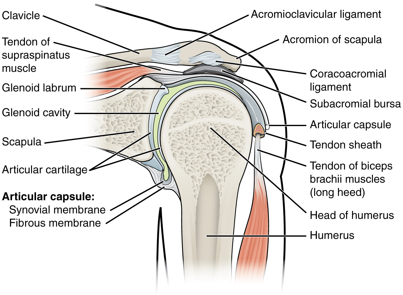
Shoulder anatomy. By OpenStax College.

Shoulder anatomy. By OpenStax College.

Differential Diagnosis

Acute Pain (Obvious/Emergent)

Acute or Chronic Pain

  • Rotator cuff injury (impingement and tear)

  • Superior labral tear from anterior to posterior (SLAP) tear

  • Thoracic outlet syndrome (less common): Compression of vessels/nerves by clavicle produces referred numbness/pain when arms are lifted to shoulder level. Arm weakness and numbness may be present on exam. Refer to physical therapy and consider surgery for refractory cases.

Chronic Pain

Shoulder Exam

  1. Assess for musculoskeletal instability from front, side and back

  2. Palpate clavicle and acromioclavicular joint

    • Assess for muscle tenderness and instability

    • Sulcus sign: Indicates shoulder instability

  3. Test range of motion:

    • Abduction (jumping jack)

    • Adduction (X across the body)

    • Elevation through forward flexion (judo chop without bending elbow)

    • Extension (swan dive)

    • External rotation (robot arms rotate outward): Decreased ROM or pain indicates adhesive capsulitis

    • Internal rotation (back scratch with the thumb)

  4. Rotator cuff

    • Empty can test (supraspinatus): Patient holds arms directly in front and pretends to empty two cans

    • Neer test (subacromial impingement): Place fully pronated arm in forced flexion

    • Hawkins-Kennedy (supraspinatus impingement syndrome): Arm bar and rotate downward

    • External lag (infraspinatus/teres minor): Resistance against external rotation and/or inability to hold arm in external rotation

    • Internal lag/Gerber Lift-off (subscapularis)

      • Resistance against internal rotation

      • Place hands behind back and lift off with/without resistance

  5. Biceps tendon and labral/SLAP tear

    • Physical exam

      • Speed test: Straight arm with palm up “asking for candy”

      • Crank test: Indicates labral/SLAP tear, especially if Speed test is positive

    • Note: Physical exam for a biceps tear can be unreliable. If suspected, obtain an MRI of the elbow for definitive diagnosis.


Elbow Pain

Adult

Medial epicondylitis pathophysiology: Tendon inflammation affecting wrsit/hand flexors. Source: Scientific Animations via Wikimedia Commons.

Medial epicondylitis pathophysiology: Tendon inflammation affecting wrsit/hand flexors. Source: Scientific Animations via Wikimedia Commons.

Mediolateral

  • Medial

    • Common etiologies

      • Medial epicondylitis (Golfer's elbow): Insidious onset in “trailing arm,” i.e. right elbow in right-handed golfer. Maximal tenderness 1 cm distal/anterior to medial epicondyle, pain with wrist flexion, decreased grip strength. Activity modification, ice, naproxen 500 mg q12h, tylenol 650 mg q6h, counterforce brace or compression sleeve, physical therapy.

      • Ulnar collateral ligament injury: UCL resists valgus stress with overhead throwing. Injuries more common in baseball, volleyball, javelin. "Popping sensation" followed by medial elbow bruising.

      • Cubital tunnel syndrome: Insidious onset. Numbness/tingling in ulnar border of forearm and hand.

    • Alarm symptoms for intra-articular pathology: Reduced elbow mobility or bony abnormalities on exam. Obtain 3-view plain elbow x-ray.

  • Lateral epicondylitis (tennis elbow)

  • Considerations if refractory to initial treatment:

    • Ulnar neuropathy

    • Radial tunnel syndrome

Anterior

  • Biceps tendinopathy: Repeated flexion/supination, e.g. dumbbell curls. NSAIDs, physical therapy. Consider corticosteroid injection in proximity to tendon.

  • Biceps rupture: Instant onset during heavy lifting. Anterior fossa tenderness, ecchymosis. Surgical repair.

Posterior

Pediatric

Hand/Wrist Pain

Unilateral

Wrist Pain Predominant

Hand Pain

  • Rheumatoid arthritis

  • Cervical radiculopathy: Neck pain with positive Spurling test and numbness pending cervical spine level affected (see dermatomal distribution).

  • Pronator syndrome (median nerve compression at the elbow): Forearm pain. Sensory loss over thenar eminence. Weakness with thumb flexion, wrist extension, and forearm pronation.

  • Ulnar compressive neuropathy: Paresthesias of the 4th and 5th digits. Positive Tinel sign and compression tests at elbow and/or wrist (Guyon canal).

Finger Pain/Immobility

  • Finger injuries including

    • Mallet fracture

    • Jersey finger

    • Skier’s thumb

  • Infections including

    • Paronychia

      • Recent trauma painful swelling along proximal/lateral nail folds. Cuticle/nail dystrophy if chronic.

      • Chlorhexidine soak, mupirocin (Bactroban), I&D if abscess present. Topical corticosteroids vs. surgery for chronic cases.

      • Counseling: Maintain dry hands.

    • Cellulitis

  • Trigger finger (stenosing flexor tenosynovitis): Due to constriction of first annular pulley overlying MCP joint

    • Initially painless, but now painful finger snapping/catching/locking

    • Naproxen 500 mg BID, triamcinolone injection q6 weeks x 2, referral to hand surgeon

  • Dupuytren's contracture

    • 50+ y/o European male with personal h/o DM, smoking, alcohol abuse, exposure to repetitive hand tasks/vibration + family history of Dupuytren's contracture. Rubs palm to straighten fingers.

    • Range of motion exercises, triamcinolone/lidocaine injection, referral to hand surgeon for collagenase injection vs. open fasciectomy

    • Contracture of 4th/5th digits, painless palmar nodules, skin tethering/puckering

Bilateral

  • Rheumatoid arthritis

  • Peripheral neuropathy: History of diabetes mellitus. Bilateral, lower extremity involvement.

  • Raynaud syndrome: Symptoms related to cold exposure, characteristic color changes of fingers (white-blue-red). Nifedipine ER 30 mg daily (MDD 120 mg daily). Follow-up every 4 weeks until symptoms are controlled: Increase daily nifedipine dose 30 mg provided BP > 120/80 mmHg.

Hip, Pelvic, Rectal Pain

Hip Pain

Adult

  • Anterior

    • Adult/Geriatric

      • Femoral neck stress fracture

      • Hip osteoarthritis +/- femoroacetabular impingement

      • Hip labral tear: Groin pain and catching/clicking sensation with activity. Positive FABER (~80%) and FADIR (~85%) tests. Obtain MRI.

      • Iliopsoas bursitis

      • Femoral hernia: Groin pain exacerbated by increased intraabdominal pressure with an upper thigh bulge on exam.

    • Hip osteonecrosis

    • Transient synovitis vs. septic arthritis

  • Posterior

    • Piriformis syndrome

    • Sacroiliac joint dysfunction

  • Lateral/Other

    • Trochanteric bursitis

    • Meralgia paresthetica

Pediatric

Pelvic Pain

Urgent/Emergent

Common

GI

Urologic

OBGYN

Rectal Pain

Visible Lesion

  • Potentially Urgent/Emergent: Trauma +/- foreign body

  • Hemorrhoid

  • Anal fissure

    • Risk factors: Hard stool/straining, pregnancy, opiate analgesia

    • Anal spasm, tearing sensation with defecation, bright red blood on stool/toilet paper

    • Treatment

      • Topical 0.4% nitroglycerin ointment administered as 1 inch paste BID PRN x 3 weeks

      • Adjunctive therapy: Docusate sodium 100 mg daily, psyllium fiber (Metamucil) 2 teaspoons in 8 oz water daily, Sitz bath daily

  • Perianal fistula, e.g. irritable bowel disease

Internal Pathology

  • Rectoanal neoplasm

  • Constipation/fecal impaction

  • Females

    • Endometriosis

    • Pelvic inflammatory disease

  • Males

    • Proctalgia fugax

    • Prostatitis

Knee Pain

Initial Considerations and Knee Exam

Red Flags

  • Acute trauma associated with

    • Severe pain, swelling, and/or instability

    • Inability to bear weight after the event

  • Signs of joint infection including

    • Fever, swelling, and overlying erythema

    • Limited range of motion

Knee Diagram.PNG

Physical Exam Maneuvers

Imaging

  • Refer for imaging after acute trauma if the patient meets one of the following criteria (Ottawa Knee Rules):

    • Age ≥ 55

    • Isolated patella tenderness

    • Tenderness at fibular head

    • Unable to flex knee to 90 degrees

    • Unable to walk ≥ 4 steps

  • Consider imaging in cases of chronic knee pain (i.e. > 6 weeks) after failure of conservative management

Diagnosis

Etiology per Mechanism and Pain Location

Trauma

  • Anterior knee pain: Patellar subluxation/dislocation

    • Adolescent with lateral patellar dislocation when the knee is held in 30 degrees of flexion, patellar effusion, and a positive apprehension test.

    • Reduction performed with the hip in flexion as lateral pressure is applied to the patella while the knee is extended. If unsuccessful, refer to orthopedics.

  • Posterior knee pain: Posterior cruciate ligament (PCL) injury

    • PCL prevents posterior displacement of tibia.

    • PCL injury: Blunt trauma to anterior tibia with knee in flexion (e.g. dashboard injury) resulting in pain with kneeling. Positive posterior “sag” sign and/or posterior drawer test. Obtain plain radiograph per Ottawa knee rules +/- MRI. Refer to orthopedics for ligament insertion avulsion and/or ≥ Grade 3 injury.

  • Mediolateral

Iliotibial band syndrome. Source: Jmarchn.

Iliotibial band syndrome. Source: Jmarchn.

Overuse Injury

  • Anterior knee pain: Avoid MRI until s/p 6 weeks of physical therapy

    • Patellofemoral pain syndrome: More common in women. Worse after prolonged sitting or when walking down stairs/hills. No associated knee effusion and positive patellar grind test.

    • Patellar tendinitis (Jumper’s Knee): Adolescent athlete involved in jumping sports who endorses infrapatellar pain with palpation of the patellar tendon, e.g. while evaluating patellar reflexes.

  • Mediolateral: Iliotibial band syndrome presenting with lateral knee pain in patients with h/o repetitive flexion (e.g. runners, cyclists). Pain present along the iliotibial band on exam.

Swelling/Effusion

  • Anterior/Anteromedial

    • Prepatellar bursitis: Anterior, unilateral knee swelling without fever, joint erythema. Initiate conservative management and consider joint aspiration.

    • Pes anserine bursitis: Tender nodule overlying the anteromedial tibia.

  • Posterior: Baker’s cyst (obtain ultrasound if suspected)

  • Diffuse Inflammatory Arthropathy

    • Unilateral or bilateral

      • Knee osteoarthritis: Age > 50 years with diffuse pain worse when initiating movement and with prolonged activity/weightbearing. Crepitus on exam; swelling/effusion may not be present.

      • Rheumatoid arthritis

    • Unilateral

      • Crystal arthropathy: Gout vs. pseudogout

      • Septic arthritis: Presenting with joint erythema, warmth, and immobility. Elevated WBC, ESR, CRP. Obtain immediate arthrocentesis with gram stain and culture if suspected.

Adolescents

  • Anterior knee pain

    • Osgood-Schlatter disease (tibial apophysitis): Age 10 to 15 years. Generally associated with a growth-spurt. Atraumatic knee pain at tibial tubercle.

    • Infrapatellar pain in adolescents: See Overuse injuries above

      • Patellofemoral syndrome

      • Patellar tendinitis (Jumper’s Knee)

  • Referred pain: Slipped Capital Femoral Epiphysis (SCFE)

    • More commonly associated with hip pain in an obese patient

    • Obtain plain radiographs and refer to orthopedics if confirmed as risk for avascular necrosis is 30% if not appropriately treated

Lower Leg/Calf Pain

Calf Pain Diagnosis.jpg

Acute/Traumatic

Subacute/Insidious

  • Muscular

    • Muscle cramp

      • Idiopathic (most common)

      • True cramp (e.g. heat-related)

      • Tetany: Thyroid disease, metabolic (e.g. hemodialysis)

      • Medication (e.g. antipsychotic)

    • Statin-induced myopathy

  • Medial tibial stress syndrome (shin splints)

  • Stress fracture

  • Infectious

Neuroendocrine

Ankle/Heel/Foot Pain

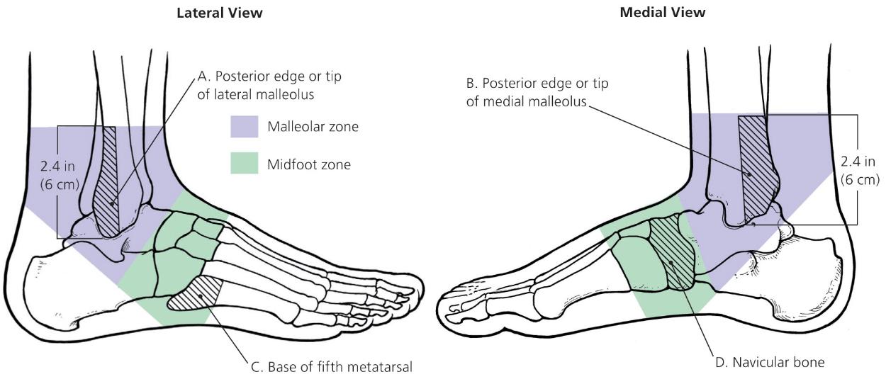
Ottawa Ankle Rules

Ankle x-ray for pain in the malleolar zone and one of the following:

  • Inability to bear weight for four steps both immediately and in ED

  • Bone tenderness at the posterior edge or tip of the lateral and/or medial malleolus

Foot x-ray for pain in the malleolar zone and one of the following

  • Inability to bear weight for four steps both immediately and in ED

  • Bone tenderness at the navicular and/or base of the fifth metatarsal

Differential Diagnosis

Plantar warts. Source: Marionette.

Plantar warts. Source: Marionette.

Heel and Ankle Pain

Adult

Plantar

  • Plantar warts: Raised skin lesions associated with pain on palpation. Lesion shaving, topical salicylic acid, imiquimod cream, and/or cryotherapy. Consider referral to podiatry for surgical excision. Duct tape is ineffective.

  • Plantar fasciitis (most common etiology): Sharp/aching anterior heel pain worse with weight bearing following rest. NSAIDs, stretches, night splinting, physical therapy. Consider corticosteroid injection, platelet rich plasma injection, extracorporeal shock wave therapy for recalcitrant cases.

  • Often confused with plantar fasciitis:

    • Heel pad syndrome: Deep mid-heel ache in worse with prolonged walking. NSAIDs, RICE, weight loss. Rigid heel cup. Consider podiatry referral.

    • Calcaneal stress fracture: Sudden increase in activity with minimalist shoes followed. Pain worse in the morning and with ambulation.

Medial

  • Maisonneuve fracture

  • Posterior tibial tendon insufficiency (PTTI)

  • Tarsal tunnel syndrome: Almost always occurs following clear foot trauma

    • Intermittent numbness and aching/burning medial/plantar foot pain worse at night and with prolonged walking

    • Pes planus, pain with ankle dorsiflexion/eversion, positive triple compression test

    • Ankle x-ray to rule out dislocation/fracture of talus, calcaneus, and/or medial malleolus; consider bilateral EMG to compare affected and unaffected foot

    • NSAIDs, shoe modifications, and/or orthotics; consider corticosteroid injection vs. nerve decompression surgery for recalcitrant cases

Eccentric calf strengthening exercises (video) for achilles tendinopathy

Eccentric calf strengthening exercises (video) for achilles tendinopathy

Lateral

Posterior

Additional Considerations

Pediatric

  • Fracture: Two-view (lateral/axial) plain film

  • Calcaneal apophysitis (Sever's Disease)

    • Elementary/adolescent athlete s/p "growth spurt"

    • Tenderness with compression of mediolateral posterior calcaneus

    • NSAID, RICE, stretching, 3 to 6 weeks rest

  • Achilles tendonitis: Tendon inflammation in adolescent athlete

Foot Pain/Discomfort

  • Acute

    • Lisfranc fracture

    • Stress fracture: Pain with walking following sudden increase in physical activity

      • Plain film and consider MRI to rule out 5th metatarsal fracture

      • Crutches (4 days) → boot (4 weeks) → re-evaluation → rigid shoe (4 weeks)

    • Acute limb ischemia (embolism)

  • Subacute and/or chronic

    • Plantar wart (see above)

    • Morton’s neuroma

    • Poikilothermia (cold feet): Raynaud’s phenomenon, atherosclerosis (peripheral arterial disease), neuropathy

Toe Pain

  • Toe fracture: Plain radiograph with anteroposterior, lateral, and oblique views

  • Gout: Excess meat/liver/beer/high-fructose corn syrup, loop/thiazide diuretic

    • Calculate acute gout diagnosis rule and CrCl per BMP. Definitive diagnosis with with aspiration.

    • Indomethacin (CrCl > 30) vs. colchicine (CrCl < 30).

Paresthesia

Peripheral neuropathy

Pruritus

Initial evaluation: CBC, CMP, TSH, HIV test

Additional evaluation: Skin scraping and/or biopsy

Skin Lesion

No Skin Lesion

Skin Lesions

Source: CNX OpenStax [CC BY 4.0 (https://creativecommons.org/licenses/by/4.0)]

Source: CNX OpenStax [CC BY 4.0 (https://creativecommons.org/licenses/by/4.0)]

Resources

Discoloration

  • Definitions: Macules and patches are circumscribed areas of discoloration that

    • Are neither raised nor depressed in relation to surrounding skin, i.e. the lesions cannot be felt when the examiners eyes are closed

    • Differ only in diameter with macules being < 1 cm and patches > 1 cm

  • Widespread skin lesions may be composed of both macules and patches

  • Remembering the terminology: Macule is derived from the Latin term for "spot." When something is spot-less, it is said to be immaculate. Patches on clothes are large and flat.

Erythematous

  • Infectious

    • Viral exanthems: Erythema infectiosum (Parvovirus B19), roseola infantum, pityriasis rosea

    • Bacterial: Cellulitis, erysipelas, cutaneous erythrasma

  • Inflammatory

    • Rosacea: Avoid facial flushing triggers, e.g. extreme temperatures, sunlight, spicy foods, alcohol, psychological stress, vasodilatory medications. Apply thin film metronidazole gel 1% daily to affected areas. Alternative agents in order of preference include topical brimonidine (Mirvaso), topical azelaic acid (Finacea), and topical ivermectin (Soolantra).

    • Henoch-Schonlein purpura (HSP - IGA vasculitis)

  • Petechiae

Hyperpigmented Lesions

Superficial

Linear

  • Fissure (crack or split)

  • Excoriation (erosion)

Deep

Plaque psoriasis. By MediaJet - Own work.

Plaque psoriasis. By MediaJet - Own work.

Lichen planus. By James Heilman, MD.

Lichen planus. By James Heilman, MD.

Epidermal thickening

  • Lichenification (exaggeration of skin lines): Atopic dermatitis

  • Scale (superficial epidermal thickening)

    • Seborrheic dermatitis: Malassezia infection generally found on scalp, face, trunk, or intertriginous zones. Treat with ketoconazole 2% shampoo (scalp) +/- topical corticosteroids.

    • Tinea (fungal infection)

      • Capitis: Griseofulvin 20 mg/kg/day in two divided doses (MDD 1,000 mg/day) x 6 weeks for children and adults.

      • Corporis, cruris, pedis, and versicolor: Start topical terbinafine 1% BID x 3 weeks. Fluconazole 10 mg/kg/day once daily dosing up to 200 mg qd (adult) x 3 weeks.

  • Crust (dried serum or exudate on skin)

    • Impetigo: Red sores typically on the face (nose/mouth) or hands that progress to honey-colored crusts. Associated with Staph aureus and/or Streptococcus pyogenes infection. Treat with topical mupirocin (Bactroban).

    • Healing vesicles: See vesicles below.

  • Plaque (elevated, solid flat lesion > 1 cm)

    • Psoriasis: Erythematous oval lesions often on extensor surfaces. Sometimes associated with pitting fingernails and asymmetric polyarthritis. Treat with topical coal tar shampoo, corticosteroids, calcipotriene. If covering > 20% of body surface or refractory, refer to dermatology for ultraviolet phototherapy, LASER excision, methotrexate, or anti-TNF therapy.

    • Lichen planus: Autoimmune response sometimes associated with medications or HCV. Pruritic, polygonal, purple plaques/papules on a white, lace-like pattern (Wickham's striae). Generally located on extremities, especially hands and wrists. Often resolves after 1-2 years. Topical and/or intralesional corticosteroids.

    • Granuloma annulare: Non-scaling annular plaque on distal extremities. Sometimes confused with tinea corporis (scaling) due to raised borders. Does not require treatment, but may respond to topical and/or intralesional corticosteroids.

Raised

Granuloma annulare. By Robert Paprstein [Copyrighted free use].

Granuloma annulare. By Robert Paprstein [Copyrighted free use].

Fluid-filled

  • Urticaria (edematous wheal)

    • Etiologies: Autoimmune response, hypersensitivity (drugs/stings/bites), physical stimuli (temperature/pressure/sunlight)

    • Treatment options: H1 blocker (e.g. cimetidine), H2 blocker (e.g. ranitidine), and doxepin (blocks H1 and H2 receptors)

  • Free fluid

    • Vesicle (< 1cm)

      • Zoster: Painful burning may precede vesicles by 4 to 5 days. Dermatomal distribution and does not cross midline. Valacyclovir 1000 mg TID x 7 days. Topical prednisone and lidocaine for acute pain. Postherpetic neuralgia treatments include capsaicin, tricyclic antidepressants, gabapentin, pregabalin, and sympathetic nerve blocks. Prevention with Shingrix (two-dose series separated by 6 months) for immune-competent adults ≥ 50 years.

      • Rhus contact dermatitis (e.g. poison ivy): Rinse contact site with water before vesicles develop. Intensely pruritic. Treat with calamine lotion, topical corticosteroids, or oral corticosteroids 1 mg/kg (MDD 60 mg) with 14 day taper. Avoid topical benzocaine (aesthetic), topical antihistamines, and antibiotics.

      • Dermatitis herpetiformis: Celiac disease

    • Bulla (> 1 cm)

      • Bullous impetigo

      • Bullous pemphigoid

      • Pemphigus vulgaris

      • Necrotizing fasciitis

    • Pustule (non-serous fluid)

      • Acne: Topical tretinoin 0.025% gel QHS → benzoyl peroxide 10% gel BID → topical erythromycin 2% gel BID → isotretinoin 1 mg/kg/day x 20 weeks

      • Pseudofolliculitis barbae: Inflammatory reaction provoked by short, tightly-curled hairs irritating skin. Discontinue shaving. Resolves within a few months.

      • Folliculitis: Superficial bacterial infection of hair follicles with S. aureus (common) or Pseudomonas (hot tub folliculitis). Apply warm compresses, avoid shaving in affected areas, and prescribe mupirocin if S. aureus is suspected. May coalesce into a carbuncle.

      • Furunculosis (boil)

      • Hidradenitis suppurativa

      • Pilonidal cyst

      • Primary bacterial abscess

Scabies Mite. By Joel Mills.

Scabies Mite. By Joel Mills.

Solid

Papule (<1 cm)

  • Malignant/Pre-malignant

  • Flesh colored

    • Warts

    • Molluscum

  • Erythematous

    • Acne: Topical tretinoin 0.025% gel QHS → benzoyl peroxide 10% gel BID → topical erythromycin 2% gel BID → isotretinoin

    • Insects and Mites

      • Bed bugs: Erythematous papule 2-5 mm in diameter with central hemorrhagic punctum. Treat bed clothes with insecticides and heat (wash in scalding water). Topical triamcinolone acetonide 0.1% + loratadine 10 mg qd for pruritus.

      • Scabies: Papules formed by a hypersensitivity reaction to mite eggs and feces. Color over bumps with a dark, felt-tipped marker and then clean with an alcohol wipe - superficial markings are removed while ink in burrows remains. Mites sometimes visualized in skin scrapings. Cover lesions with permethrin 5% cream daily and wait 10 hours before washing location. Ivermectin (Soolantra) 1% cream for recalcitrant cases.

    • Lichen planus: See plaques above

  • Hyperpigmented

    • Seborrheic keratosis

    • Dermatofibroma

Nodular (> 1 cm) and/or cystic (membranous sac)

  • Lipoma

  • Epidermal inclusion cyst

  • Mucoid cyst

  • Ganglion cyst

  • Hypertrophic scars

Syncope

Differential Diagnosis

Syncope.jpg
  • Neurally mediated (45% of cases): Increased parasympathetic/vagal tone → bradycardia and hypotension

    • Vasovagal: Syncope occurred in a warm/crowded space after prolonged standing and was associated with emotion/fear/pain. Patient reports associated nausea and bystanders noted transient diaphoresis.

    • Situational: New onset syncope occurred during episode of coughing while voiding after a meal

    • Consider seizure (eyes open) and psychogenic syncope (eyes closed with rapid, complete recovery)

  • Cardiac (20% of cases): Generally considered a high risk patient (see below)

    • Arrhythmia (most common): Elderly patient with personal h/o atrial fibrillation/flutter and family h/o sudden, unexplained death presents with new onset palpitations during exercise and abnormal EKG. Treatment per arrhythmia.

    • Structural cardiac abnormality/cardiomyopathy: Elderly patient with h/o valvular and infiltrative (sarcoidosis, hemochromatosis, amyloidosis) heart disease presents chest pain at rest, syncope during exercise. Evidence of heart failure on physical exam and PR interval > 200 ms (heart block) on EKG.

    • Hypertrophic cardiomyopathy: Pediatric patient with family h/o sudden cardiac death presents with new onset syncope that occured while exercising in hot weather. Start nadolol; transition to verapamil if ineffective.

  • Orthostatic hypotension (10% of cases): Decrease of ≥ 20 mmHg systolic or ≥ 10 mmHg diastolic within 3 minutes of moving from supine to upright position

    • Patient with h/o autonomic dysfunction due to alcoholism, DM, Parkinson disease, multiple sclerosis presents with syncope during postural change. Recent, acute volume loss due to dehydration, hemorrhage. Consumes low-salt diet.

    • Recent changes in medications: Alpha blockers (tamsulosin), beta-blockers (metoprolol), calcium channel blockers (amlodipine), diuretics (furosemide), phosphodiesterase inhibitors (e.g. sildenafil), SSRIs

    • Tachycardia and positive orthostatic vital signs on exam

Evaluation and Treatment

Obtain history with focus on precipitating events, h/o cardiac disease, and clinical features above

  • Obtain orthostatic vital signs

  • Obtain CBC, CMP; consider BNP if evidence of heart failure

  • Evaluate EKG for prolonged PR interval, arrhythmia

  • Specific etiologies

    • Neurogenic syncope

      • Vasovagal/situational hypotension: IV hydration, patient education. Consider tilt table test to confirm bradycardia/hypotension with change in position.

      • Event not explained by vasovagal or situational syncope: Consider EMG, psychiatry consult

    • Unexplained and/or suspected cardiac syncope

      • High risk: Telemetry, echocardiogram. Consider Holter monitor/loop recorder, cardiac stress test.

      • Low risk or negative hospital workup: Consider Holter monitor/loop recorder to detect arrhythmia

    • Orthostatic hypotension: Modify risk factors; if refractory, consider starting midodrine, fludrocortisone, or droxidopa

  • Risk stratification

    • Admit to hospital if patient is high risk due to any of the following: Age > 50 years, syncope with exertion, personal h/o heart disease, familial h/o sudden death, systolic < 90 mmHg, arrhythmia on EKG

    • Risk of adverse event within 30 days calculated per Canadian Syncope Risk Score

Urinary Retention and Incontinence

Retention

General

Etiology

  • Outflow obstruction

  • Neurologic

  • Iatrogenic: Medications commonly associated with retention include

    • Anticholinergics: Scopolamine, oxybutynin (bladder antispasmodic), dicyclomine (Bentyl), benztropine

    • Antihistamines: Diphenhydramine, hydroxyzine

    • Muscle relaxants: Cyclobenzaprine (Flexeril)

    • Neurologic/Psychiatric

      • Tricyclic antidepressants: Amitriptyline, nortriptyline

      • Antiparkinsonian: Amantadine, levodopa (Sinamet)

      • Antipsychotics: Haloperidol, chlorpromazine, fluphenazine (Prolixin), prochlorperazine (Compazine)

    • Hormonal: Estrogen, progesterone, testosterone

Incontinence

General Incontinence Interview

3 Question Incontinence.PNG
  • Perform 3 Question Incontinence Questionnaire

  • Rule out red flags including hematuria, obstructive symptoms, recurrent UTIs

  • Rule out reversible causes (DIAPPERS)

    • Delirium

    • Infection (acute UTI)

    • Atrophic vaginitis

    • Pharmaceuticals, e.g.

      • Antihistamine/anticholinergic (retention → overflow incontinence)

      • Antihypertensive: Diuretics, ACE inhibitors, Calcium channel blockers

      • Pain medications: COX-2 NSAIDs, muscle relaxants, opioids

    • Psychological disorder, especially depression

    • Excessive urine output (e.g. hyperglycemia)

    • Reduced mobility (i.e. functional incontinence)

    • Stool impaction (constipation → retention → overflow incontinence)

  • Explore effect of symptoms on patient’s quality of life

  • Review 3 day voiding diary

Differential Diagnosis

Failure to Gain Weight

Fever

Adult

Common Infectious Etiologies

Fever of Unknown Origin Considerations

Hot Flashes and Flushing

  • Menopause: 45+ y/o F with 12+ months amenorrhea and no alternative biophysiologic explanation

    • Hot flashes, irregular menses, vaginal dryness, dyspareunia, sleep disturbances, depression/mood change

    • Labs

      • TSH if concern for hyperthyroidism

      • Age < 45 with amenorrhea: Obtain hCG, prolactin, TSH, FSH

    • Counseling: Expectations per STRAW staging system

  • Emotional flushing (embarrassment)

  • Flushing due to medications/food/alcohol

  • Rare: Paraneoplastic syndromes

    • Renal cell carcinoma

    • Endocrine tumors

      • Medullary thyroid carcinoma of the thyroid

      • Pheochromocytoma

      • Pancreatic islet-cell tumors

      • Carcinoid syndrome

Additional reading: Differential Diagnosis of Hot Flashes

Pediatric

Etiology

Fever of Unknown Origin

Fever of unknown origin definition: Present on most days for at least 3 weeks with unclear etiology after 1 week of intense investigation

  • Etiology

    • Common conditions with atypical presentation (most common)

    • Infection

    • Neoplastic disorders

    • Autoimmune disease

    • Vasculitis: Henoch-Schönlein purpura, Kawasaki disease, Behçet's disease

  • Diagnosis

    • Aerobic and anaerobic blood cultures, CBC with manual differential, peripheral blood smear, CMP, ESR, CRP

    • Urinalysis with culture

    • CXR

Hypertension


Hyponatremia

Differential

Sodium Correction

  • Methods

    • Initial

      • Stop medications that may be contributing

      • Fluid intake restriction

    • 3% NaCl

  • Limit correction to < 12 mmol/L in 24 hours and < 18 mmol/L in 48 hours

Microscopic Hematuria

Approach

Microscopic hematuria definition: Appropriately collected void with

  • 3 or more RBCs per high-powered field

  • No evidence of infection

Evaluation

  • “Dirty sample,” e.g. > 3 bacteria species and/or multiple squamous cells: Repeat U/A

  • Urinalysis suggestive of UTI, e.g. positive leukocyte esterase +/- nitrites: Treat UTI

  • Patient menstruating or other reasonable explanation (e.g. calcium oxalate crystals on U/A, recent history of vigorous exercise/trauma): Repeat U/A in 6 weeks

  • Age > 35 years: Refer to urology for cystoscopy if any of the following apply

    • No alternative explanation, e.g. exercise, trauma, illness, infection

    • Patient has any risk factors for urinary malignancy, e.g. excessive NSAID use, smoking, exposure to chemical dyes

  • Consider referral to nephrology for any the following findings on urinalysis:

    • Proteinuria in the setting of DM, HTN, and/or elevated creatinine levels

    • Presents of albumin and/or cellular casts

AAFP Algorithm for Asymptomatic Microhematuria (2013)

Differential

Pyuria

Hyperthyroidism

Hyperthyroid workup.PNG

Differential Diagnosis

  • Sub-clinical

  • Graves disease

    • Diplopia, blurred vision, exophthalmos, goiter

    • Positive TPO antibodies and homogeneous radioactive iodine uptake

    • Methimazole vs. thyroidectomy vs. radioiodine ablation

  • Toxic multinodular goiter

    • Elderly patient with iodine deficient diet

    • Radioactive iodine scan: Multiple nodules with high uptake

    • Treatment: Propranolol, methimazole

  • Transient (painless) thyroiditis

    • Thyroiditis symptoms with normal physical exam

    • TSH persistently between 0.1 and 0.4 mIU/L

    • Rule out iatrogenic etiology (amiodarone, lithium)

  • Sub-acute (De Quervain) thyroiditis

    • Painful thyroid s/p febrile illness +/- rash

    • TSH between 0.1 and 0.4 mIU/L

    • Naproxen for pain and repeat testing at 6 weeks

Alternative Diagnosis: See thyroid nodule evaluation

Hypocalcemia

Hypercalcemia

Additional Resources: Clinical Problem Solvers Schema

Elevated PTH

Normal PTH

  • Hypercalcemia associated with

    • Malignancy

    • Granulomatous disease

    • Thiazide diuretics

    • Thyrotoxicosis

    • Immobilization

Electrocardiogram

Reading Checklist with Normal Findings

  • Calibration: 1.0 mV vertical box inscription (standard = 10 mm)

  • Heart rate calculation methods (choose one)

    • Regular or irregular rhythm: (Number of QRS complexes in 10-second rhythm strip)*6

    • Regular rhythm

      • Rate = 60/RR interval

      • Example when RR interval is one large box (0.2 seconds): 60/0.2 = 300 BPM

    • Regular rhythm: Estimation per number of large boxes

      • 1 large box = 300 beats/min (duration = 0.2 sec)

      • 2 large boxes = 150 beats/min (duration = 0.4 sec)

      • 3 large boxes = 100 beats/min (duration = 0.6 sec)

      • 4 large boxes = 75 beats/min (duration = 0.8 sec)

      • 5 large boxes = 60 beats/min (duration = 1.0 sec)

  • Rhythm

    • All QRS complexes preceded by normal P

    • No ectopy, i.e. no early beats

  • Morphology

    • PR interval 0.1 to 0.2 seconds (1/2 to 1 large box) and followed by QRS

    • QRS

      • Axis: Upright in BOTH lead I and aVF (-30 to +100)

      • Interval < 0.12 ms (3 small boxes)

    • QT interval < 440 ms (2 large boxes)

  • Hypertrophy: No RAD or LAD

    • R wave < 35 mm in V5 or V6 + SV1 or V2

    • R aVL < 11 mm

  • Infarction: No Q-waves

  • Repolarization

    • Frontal: T upright where QRS upright

    • Precordial: T always upright in V4-V6

    • QT < 50% RR interval

Bradycardia

Tachycardia

  • Sinus tachycardia: P wave preceding each QRS interval with a normal P-wave axis

  • Atrial tachyarrhythmias

  • Atrial fibrillation

    • Acute

    • Chronic

  • Atrial flutter


Miscellaneous Differentials

Lesions of the Hand and Wrist

Differential

  • Soft tissue

    • Acute

    • Subacute/chronic

      • Ganglion cyst (most common): Smooth, firm mass generally found at wrist, flexor tendon sheath of digits, distal interphalangeal joint. Trial of aspiration if painful. Definitive treatment with excision.

      • Giant cell tumor of tendon sheath (second most common): Slow-growing, firm, nontender fixed multinodular mass generally found on 1st, 2nd or 3rd digit. Hand x-ray appears normal or shows soft tissue mass. Refer for surgical resection.

      • Epidermal inclusion cyst (third most common): Penetrating trauma induces keratin production. Slow growing, firm, painless mass generally found on tactile surface of digits. Hand x-ray if diagnosis in doubt. Refer for surgical excision.

      • Foreign body granuloma: Cellular proliferation around foreign body. Similar clinical appearance to epidermal inclusion cyst. Obtain hand x-ray. Surgical excision if symptomatic.

      • Dupuytren contracture with fibromatosis: Palmar fascia fibromatosis. Non-tender palmar mass with dimpling. Consider biopsy if diagnosis is in doubt.

      • Hemangioma: Proliferating blood vessels generally found during first few years of life. May grow rapidly for up to 1 year. Seventy percent involute by age 7 years. Infection and ulceration may occur and are treated with wound compression, wound care, and antibiotics. Surgical referral for failure to involute by age 7 years.

  • Cartilage…

  • Bone

    • Enchondroma: Most common primary bone tumor. Cartilage overgrowth that generally affects phalanges. Hand x-ray shows well circumscribed, radiolucent lesion often with punctate calcifications. Refer for surgical resection.

    • Intraosseous ganglion: Most common cystic lesion of bone. Benign, locally aggressive, generally affects carpal bones. Obtain hand x-ray. Refer for surgical curettage and bone grafting if symptomatic.

    • Brown tumor: Associated with primary and secondary hyperparathyroidism (CKD). Increased PTH induces osteoclast activity and bone remodeling. Most common in distal phalanges. Hand x-ray shows endosteal resorption and scalloping.

  • Latent mass: Remain unchanged or heal spontaneously

    • Vascular aneurysms: Generally form following trauma. Painless unless compressing surrounding nerves.

  • Active masses: Grow within anatomic boundary and generally require surgery

    • Lipoma

  • Aggressive masses: Grow beyond anatomic boundaries

    • Giant cell tumor of tendon sheath: Second most common hand mass

General history

  • Lesion: Rate of growth, changes in consistency/color

  • ROS: Pain, paresthesias, motor dysfunction

  • Red flags: Rapid growth, night pain, sudden increase in pain

  • PMH: Recent trauma, renal disease, parathyroid disease, prior malignancy

  • PE: Document overlying skin color, mass mobility, mass consistency

  • Obtain hand x-ray

Source: Tumorlike Lesions and Benign Tumors of the Hand and Wrist


Wilderness Medicine

  • Protocols: AVPU, SIPSO, and MARCH

  • Anaphylaxis

  • Burns

  • Animals

    • Bites/stings/envenomation

    • Arthropod-borne illness

  • Exposure/environmental

    • Hypothermia/Hyperthermia

    • Submersion injuries

    • High altitude illness

  • Trauma

    • Wound management

    • Patient patient packaging/evaluation

      • MSK injuries and splinting

      • C-spine stabilization

      • Evacuation

  • Professional Opportunities

    • High adventure sports medicine

    • Dive medicine

    • Travel/expedition medicine

    • Tactical and disaster medicine

Wilderness Medicine

Acute mountain sickness

  • Unacclimated pt at > 2500 m with at least one of the following symptoms: Anorexia, nausea, vomiting, insomnia, dizziness, or fatigue

  • Treatment options: Dexamethasone, acetazolamide

  • High altitude cerebral edema diagnosed if AMS or ataxia develops (administer supplemental O2 and descend 500 to 1000 m)

High altitude pulmonary edema: Nifedipine